

 Print
Print facebook
 twitter
 Linkedin
 google+

Introduction
Cephalopelvic disproportion (CPD) is a clinical diagnosis made during labor when the progress of labor is not satisfactory, resulting in the performance of a cesarean section. CPD remains one of the commonest indications for the performance of an emergency cesarean section. Several studies, using a variety of clinical assessment modalities, have been published in an attempt to predict which women have CPD before the onset of labor [1-4]. No single modality or assessment protocol at present has been able to reliably predict successful vaginal delivery before labor [2]. Radiographic methods for the prediction of CPD have become less popular, as they have been shown to have poor predictive value [3,4]. Alternative modalities such as computed tomography and magnetic resonance image, although possible, are limited by their use of high doses of radiation, their lack of portability, and cost [5-7].
Digital vaginal examination during labor is still the gold standard used to diagnose whether labor is progressing [8]; however, it is has been shown that digital vaginal examination is highly operator-dependent and therefore unreliable [9,10]. Intrapartum ultrasound has been suggested as an alternative to digital examination, as this modality could provide a more objective assessment of the labor process by allowing a direct assessment of the fetal location, the size of the fetal presenting part, fetal descent, and maternal pelvic size [11-14]. Current proposals for the ultrasonographic assessment of labor have focused on describing the relative position of the fetal head to the pubic symphysis; few have considered whether ultrasonography could be used to measure pelvic tissue thickness and whether soft tissue thickness is related to the mode of delivery. The retropubic tissue thickness (RTT), corresponding to the soft tissue between the bony maternal pelvis and fetal skull, is a parameter that could be useful in this context. It corresponds to the space between the pelvic bone and fetal skull as the fetal head descends, and could be measured using ultrasonography.
In the present study, we first describe a new and simple ultrasonographic method that could be used to measure the RTT, then present an evaluation of the measurementŌĆÖs reproducibility, and lastly evaluate the potential clinical association between RTT and mode of delivery.
Materials and Methods
Nulliparous women between October 2014 and August 2015 who had a singleton pregnancy with a fetal cephalic presentation, who were admitted with regular uterine contractions, a fully effaced cervix, and a cervical dilatation of 2-3 cm on digital examination, or who were scheduled to have an induction after 37 weeks of gestation, were invited to participate in the study. Women who gave written consent underwent serial ultrasound scans between uterine contractions every 1 hour until they delivered. At each time point, a 3-dimensional (3D) volume was acquired of the fetal skull and pubic symphysis by a transperineal scan as described in detail in the literature [11-13]. In brief, the ultrasound probe was covered with a protective glove and placed longitudinally at the labia without exerting any pressure.
A 3D volume was acquired between uterine contractions and stored after the long axis of the pubic symphysis and the outline of the fetal skull had been visualized. The 3D capability was then engaged by switching to the volume mode. The volume box was placed over the entre image covering the pubic symphysis. All scans were performed by one of two sonographers, each with over 6 years of clinical experience performing fetal morphology scans, using a GE Voluson i (General Electric Medical Systems, Zipf, Austria) ultrasound machine using a RAB2-5-D H48651MN (1-4 MHz) volumetric probe. Post-delivery analysis of the acquired ultrasound volume was performed using 4D View (ver. 14, General Electric Medical Systems).
All clinicians remained unware of the RTT measurements, and labor was managed in accordance with our internally published departmental standardized protocols. The study was approved by the Chinese University of Hong Kong Clinical Research Ethics Committee.
Retropubic Tissue Thickness Measurement
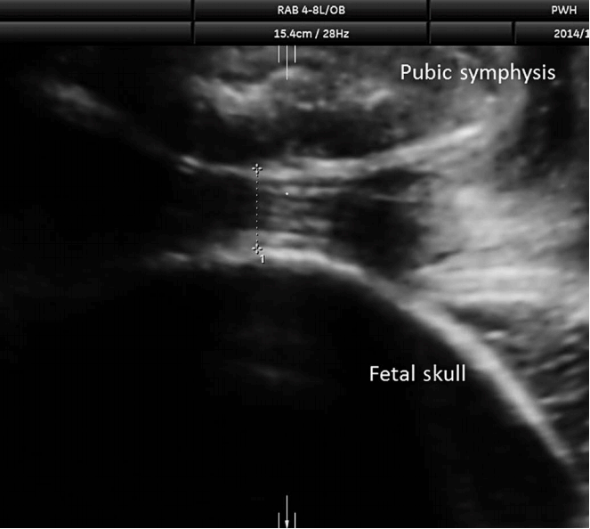
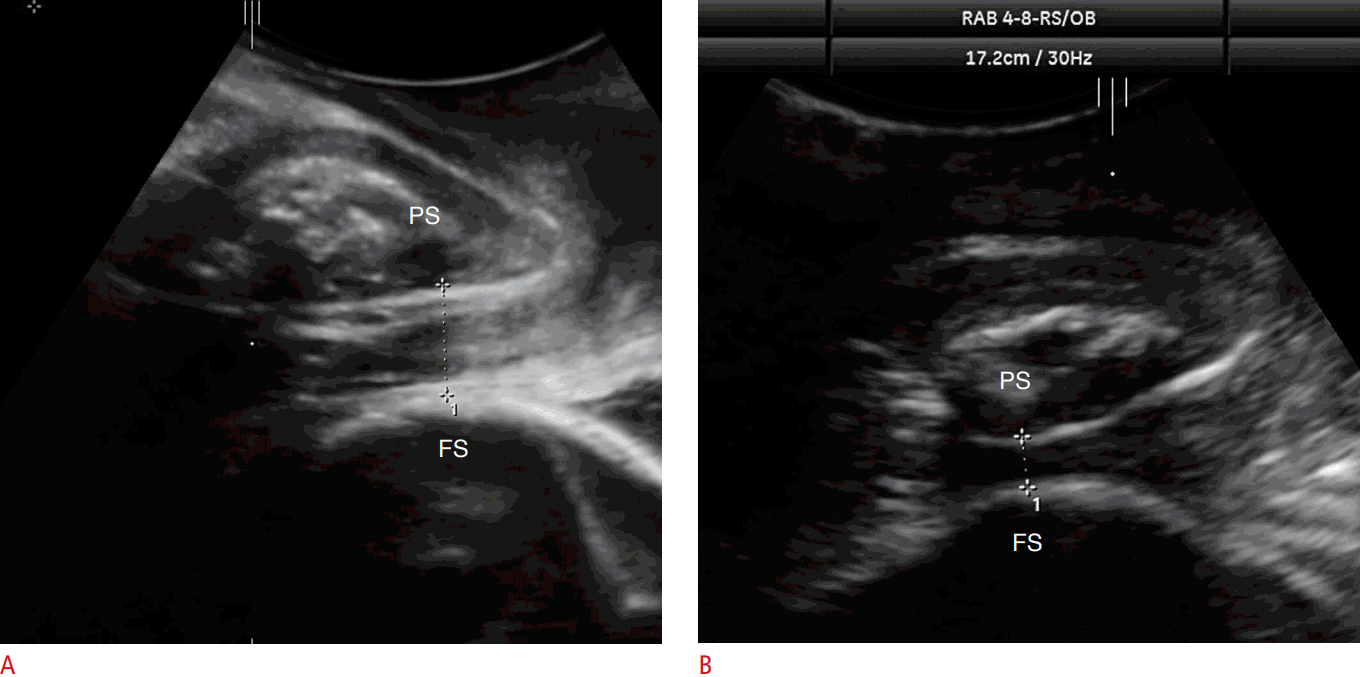
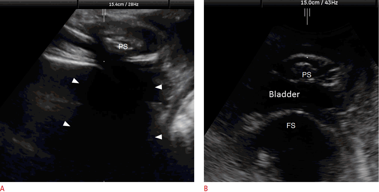
Retropubic tissue thickness was retrospectively measured by first visualizing the pubic symphysis and the fetal skull on the sagittal ultrasound plane. Images were then magnified (├Ś5) in the azimuth plane. The RTT was then measured by determining the shortest vertical distance between the outer capsule of the pubic symphysis and the fetal skull. Measurements were taken by placing the inner border of the horizontal line of the calipers on the hypoechogenic area of the pubic symphysis just above the line that defines the outer capsule of the pubic symphysis and the outer border of the fetal skull, as shown in Figs. 1 and 2. During measurement, care was taken to ensure that bladder tissue was not present between the fetal skull and the pubic symphysis (Fig. 3). In addition, the border of the pubic symphysis and the outline of the fetal skull were required to be clearly visible, as shown in Fig. 2.
Intrasonographer and Intersonographer Variation in Retropubic Tissue Thickness
Three-dimensional volumes were retrieved, and the RTT was measured as described above by three independent sonographers (A, B, and C). Sonographer A was an operator experienced in performing intrapartum ultrasound scans, whereas sonographers B and C had performed antenatal ultrasound examinations for more than 5 years. Intrasonographer repeatability was assessed by calculating the repeatability coefficient (r) defined as 1.96├Ś 2 3 S 2 / n
Retropubic Tissue Thickness and Mode of Delivery
The 3D volumes acquired at 1-hour intervals during labor were retrieved, and the RTT and angle of projection (AoP) were measured by sonographer A. The AoP was determined by methods described in the literature [12] and was defined as the angle between the long axis of the pubic symphysis and a line extending from the inferior border of the pubic symphysis to the tangent of the outline of fetal skull. The RTT before the AoP exceeded 120┬░ was documented and used to assess the association between the RTT and womenŌĆÖs eventual mode of delivery. An AoP <120┬░ was used as it has been reported that laboring women who can attain an AoP greater than this level should be able to deliver vaginally [16]. Women were stratified according to their eventual mode of delivery (vaginal, instrumental, or cesarean). Continuous variables are presented as mean┬▒SD or median and interquartile range, while qualitative variables are presented as absolute frequency. Comparisons between categorical variables were tested by the use of contingency tables using the Fisher exact test. Comparisons between normally distributed continuous variables were performed using one-way analysis of variance, and if justified, by a between-group comparison using the Fisher least significant difference with statistical significance adjusted for multiple comparison using a Bonferroni correction.
All statistical analyses were performed using SPSS ver. 22.0 (IBM Corp., Armonk, NY, USA). P-values <0.05 were considered to indicate statistical significance.
Results
A total of 129 women participated in the study. The RTT could not be determined in 24 women (18.6%), as the border of the pubic symphysis or the fetal skull was not well visualized in eight cases (6.2%), and bladder tissue was seen between the fetal skull and the pubic symphysis in 16 cases (12.4%) (Fig. 3). Table 1 summarizes the patient characteristics and mean RTT measurements by eventual mode of delivery. Analysis of variance indicated that RTT was significantly associated with the overall mode of delivery (P=0.011) and the birth weight of the baby (P<0.001). A between-group comparison indicated that RTT was significantly greater in women who had a spontaneous vaginal delivery than in those who had a cesarean delivery (P=0.008). There were no statistically significant differences in RTT between patients who had a normal vaginal delivery and patients who had an instrumental delivery (P=0.990), or between those who had an instrumental delivery and those who had a cesarean delivery after correcting for multiple significance testing (P=0.120). The Mahalanobis distance, the difference in means over the standard deviation, between those who had a vaginal or cesarean delivery was 0.69 (0.22/0.32). Patients who delivered by cesarean section (P=0.001) or by instrumental delivery (P=0.001) had a greater birth weight than patients who delivered by spontaneous vaginal delivery.
Tables 2 and 3 report the measurement ranges and the interobserver assessments of RTT in the first 35 women who had a measureable RTT. The measured RTT ranged from 0.45 to 1.89 cm. The intersonographer bias ranged from -0.05 to 0.04 cm. The repeatability coefficient of operator A was 1.2 mm. The overall interoperator ICC of the three operators was 0.97 (95% confidence interval [CI], 0.95 to 0.98).
Discussion
CPD remains a clinical diagnosis during labor based on indirect evidence, such as arrest of cervical dilatation and molding of the fetal head. However, there are limitations in these parameters, and they are well known to lack objectivity. In contrast, our study showed that measuring the RTT was a reliable and reproducible method of assessing CPD during labor.
Our study is the first to develop an ultrasound-based objective method to measure the relative spaces of the birth canal during labor by directly measuring the thickness of the vaginal tissue of the mid-pelvis after the fetal head is engaged. This may reflect the true pathophysiology of CPD. RTT is a direct measurement of the soft tissue between the bony maternal pelvis and fetal skull during labor. CPD occurs when the dimension of the presenting part (e.g., vertex in an occipital anterior fetal head position) is larger than the dimension of the maternal pelvis at the narrowest point. Since fetal head molding can occur during labor and soft tissue in the birth canal can be compressed by the fetal head to a certain extent, RTT measurements analyze these two dynamic factors, which cannot be achieved by traditional pelvimetry. Our data suggest that RTT is a discriminatory marker, and that by measuring RTT during labor, it will be possible to identify patients who ultimately need a cesarean section, because they have a lower RTT due to the presence of less space between the maternal bony pelvis and the fetal skull. This finding is encouraging but not conclusive. A prospective study is necessary to confirm our findings. Our preliminary data indicated that RTT assessment, although discriminatory, would probably need to be combined with other assessments, as a standalone assessment would require the Mahalanobis distance to be at least over 3. The Mahalanobis distance for RTT in our study was 0.73, which is on par with the Mahalanobis distance of biochemical markers such as ╬▒-fetoprotein and unconjugated estriol currently used to estimate individual womenŌĆÖs risk of Down syndrome in the second trimester.
Because measuring RTT is a totally different modality for evaluating the relative spaces of the birth canal in laboring patients, we suggest that this could be a complementary assessment tool to digital vaginal examination and other intrapartum ultrasound parameters (e.g., AoP, head progression distance, and head-symphyseal distance).
Our results show that this new proposed method for assessing CPD was highly reproducible and repeatable. The repeatability coefficient of 1.2 mm indicates that the absolute difference between any two future measurements by the operator would be no greater than 1.2 mm on 95% of occasions. The interoperator ICC was 0.97 (95% CI, 0.95 to 0.98), which is higher than the level usually considered acceptable.
A limitation of our study is the retrospective nature of the data analysis. The same volume was assessed by three different operators to determine the intraobserver error and interobserver error. Whether or not such a high reproducibility of the method can be confirmed in a prospective study is yet to be determined. Preferably, the three operators should acquire the volume independently. Nevertheless, as a proof-of concept trial, we have shown that RTT is a new ultrasound parameter for evaluating CPD.
In our study, the RTT measurements were obtained from 3D volumes acquired in a prospective study. All RTT measurements are taken from the azimuth plane from a 3D volume, which makes it reasonable to think that RTT measurements should be possible in 2-dimensional (2D) ultrasound images as well. We believe that measurements of RTT on 2D images and 3D volumes will yield similar results, although a future study would be required to confirm this by directly comparing 2D measurements of RTT and the offline analysis of 3D volumes.
In conclusion, with intrapartum transperineal ultrasound, it is feasible to measure RTT, with satisfactory intraoperator repeatability and interoperator reproducibility. This easy-to-learn technique extends the possibilities of assessing CPD in an objective way. Further prospective studies will be required to estimate the sensitivity and specificity of RTT as a method of distinguishing between women who have a vaginal or cesarean delivery, either by itself or in combination with other potential physical examinations and clinical history within a multi-assessment model.
 Download Citation
 PDF Links
 PubReader
 ePub Link
 Full text via DOI
 Full text via PMC


