AbstractPurpose:This study was designed to determine the rate of ductal carcinoma in situ (DCIS)underestimation diagnosed after an ultrasound-guided 14-gauge core needle biopsy (US-14G-CNB) of breast masses and to compare the clinical and imaging characteristics between trueDCIS and underestimated DCIS identified following surgical excision.
Methods:Among 3,124 US-14G-CNBs performed for breast masses, 69 lesions in 60 patients were pathologically-determined to be pure DCIS. We classified these patients according to the final pathology after surgical excision as those with invasive ductal carcinoma (underestimated group) and those with DCIS (non-underestimated group). We retrospectively reviewed and compared the clinical and imaging characteristics between the two groups.
Results:Of the 69 lesions, 21 were shown after surgery to be invasive carcinomas; the rateof DCIS underestimation was 30.4%. There were no statistically significant differences withrespect to the clinical symptoms, age, lesion size, mammographic findings, and ultrasonographic findings except for the presence of abnormal axillary lymph nodes as detected on ultrasound. The lesions in 2 patients in the non-underestimated group (2/41, 4.9%) and 5 patients in the underestimated group (5/19, 26.3%) were associated with abnormal lymph nodes on axillary ultrasound, and the presence of abnormal axillary lymph nodes on ultrasound was tatistically significant (P=0.016).
Ultrasound-guided core needle biopsy (US-CNB) is an invaluable tool for the diagnosis of breast lesions and has many advantages compared to stereotactic biopsies, including a lack of ionizing radiation exposure, increased patient comfort, lower cost, reduced procedure time, and real-time visualization of needle placement [1-3]. However, the possibility of histologic underestimation of lesions and false negative results by core needle biopsy has been unavoidable despite improvements in biopsy devices and efforts to reduce missed breast cancers, including imaging-pathologic correlation with repeat biopsy. Ductal carcinoma in situ (DCIS) underestimation occurs when a lesion is determined to be DCIS after a percutaneous breast biopsy and is subsequently shown to be an invasive carcinoma following surgical excision. The rate of underestimation is likely due to sampling error in lesions that contain both DCIS and invasive cancer. On mammography, DCIS is typically depicted as calcifications, although a lesion may also appear as a non-calcified mass. In a review of the literature, most prior studies of DCIS underestimation have been performed using stereotactic devices, including cases with directional vacuumassisted biopsy and those with automated large core needle biopsy, and have reported variable underestimation rates ranging from 5%-44% [4-8]. However, DCIS underestimation after a US-CNB of a breast mass has not been thoroughly evaluated, and studies on the ultrasonographic factors related to DCIS underestimation after a US-CNB are also very rare [9]. The purpose of this study was to determine the rate of DCIS underestimation following US-guided 14-gauge CNB (US-14G-CNB) of a breast mass and to compare the clinical and imaging characteristics between true DCIS lesions and underestimated DCIS lesions identified following surgical excision.
Our Institutional Review Board approved this study. Informed consent was not required from patients for this retrospective analysis.
Between July 2005 and July 2007, 3,124 US-14G-CNBs of breast masses were performed at the breast imaging center at our institution. Among the lesions, 78 lesions (2.5%) were pathologicallydetermined to be DCIS. The inclusion criterion for this study was a histopathologically-proven pure DCIS without signs of microinvasion or invasive cancer from a core biopsy specimen as determined by the use of light microscopy. DCIS with microinvasion was defined as tumor cells, singly or in clusters, that had infiltrated the periductal stroma or were seen as a projection of neoplastic cells through a disrupted basement membrane in continuity with the DCIS, measuring ≤1 mm along the greatest dimension [10]. Excluding lesions with microinvasions (n=9), 69 pure DCIS lesions from 60 patients were identified that manifested as identifiable masses with or without calcifications as depicted on ultrasonography and were included in the study population. Six patients had two separate lesions and one patient had four separate lesions.
All of the patients underwent a clinical breast examination, mammography, and breast ultrasonography. The mammograms were performed using a Lorad/Hologic Selenia Full Field Digital Mammography system (Lorad/Hologic, Danbury, CT, USA). Standard craniocaudal and mediolateral oblique views were routinely obtained, and additional mammographic views were used as needed. Breast ultrasonography was performed by one of five radiologists with a specialty in breast imaging, and high-resolution ultrasonography units with a 7-12 MHz linear array transducer (ATL HDI 5000 or 3000; iU22; Philips Medical System, Bothell, WA, USA) were used for breast ultrasonography.
Two radiologists retrospectively reviewed the mammographic and ultrasonographic findings of the biopsied lesions resulting in DCIS by consensus. The mammographic characteristics of the lesions were classified as negative, calcifications only, a mass, a mass with calcifications, asymmetry, and asymmetry with calcifications. The ultrasonographic characteristics of size, shape, orientation, margin, lesion boundary, and echogenicity of nodules according to the American College of Radiology Breast Imaging Reporting and Data System (BI-RADS) lexicon were reviewed retrospectively [11]. The lesion size was measured according to the maximum lesion diameter as measured on ultrasonography. When performing breast ultrasonography, the bilateral axillary regions were also assessed for the presence of abnormal lymph nodes. An abnormal lymph node was defined as a lymph node with an eccentric or irregular cortical thickening (usually >3 mm) irrespective of lymph node size, round shape (short-to-long diameter ratio >0.5), change in internal echogenicity (hyperechoic, cystic change, or calcification), or absent or compressed echogenic hilum in accordance with previous reports [12-14]. In the ultrasonography evaluation of the axillary lymph nodes as normal or abnormal, we did not use a size as a definite diagnostic criterion for this study. Although larger nodes tend to have a higher incidence of malignancy, reactive nodes can be as large as metastatic nodes. Thus, nodal size alone cannot be used to distinguish reactive nodes from metastatic lymph nodes.
The clinical records for the 60 patients were reviewed to determine their age and symptoms at the time of presentation.
A US-14G-CNB was performed using a free hand technique and a high-resolution ultrasonography unit with a 7-12 MHz linear array transducer (ATL HDI 5000 or 3000; iU22). All procedures were performed using an automated gun (Pro-Mag 2.2; Manan Medical Products, Northbrook, IL, USA) and 14-gauge Tru-Cut needles with a 22 mm throw (SACN Biopsy Needle; Medical Device Technologies, Gainesville, FL, USA). One of five radiologists specializing in breast imaging performed all of the biopsies. Prior to biopsy, a breast ultrasonography (including the bilateral axillae) was meticulously performed. A minimum of five biopsy samples were obtained with additional samples collected at the discretion of the radiologist. Informed consent was obtained from each patient undergoing a biopsy. The pathologic results of the US-14G-CNBs for each case were retrospectively reviewed with the final pathology findings as determined after breast surgery.
The results of the US-14G-CNBs were correlated with the subsequent surgical (conserving surgery or mastectomy) histologic findings. Axillary lymph node status was determined after a sentinel lymph node biopsy or axillary lymph node dissection. The rate of underestimation was defined as a diagnosis of DCIS after a US-14GCNB with a pathologic diagnosis of invasive carcinoma following surgery. The patients were classified into either the underestimated or non-underestimated group. The underestimated group was defined as cases diagnosed as DCIS after a US-14G-CNB but later determined to be invasive ductal carcinoma (IDC) following surgical excision. The non-underestimated group consisted of cases diagnosed as DCIS after a US-14G-CNB and determined not to have invasive cancer following surgical excision. We evaluated the differences between the underestimated and non-underestimated groups in terms of age, clinical symptoms, mammographic findings and ultrasonographic characteristics, including axillary findings. Tests for statistical significance were performed using SPSS ver. 12.0 (SPSS Inc., Chicago, IL, USA). A P<0.05 was considered significant. Statistical comparisons were performed using the chi-squared test (Fisher exact test) for categorical variables and the independent t-test for continuous variables. Confidence intervals were calculated according to the formula developed by Berry [15].
All 60 patients were women (age range, 24 to 88 years; mean age, 47.5±11.3 years). Of the 69 lesions diagnosed as DCIS after US- 14G-CNB, invasive carcinoma was diagnosed following surgical excision in 21 lesions from 19 patients (the underestimated group). Thus, the DCIS underestimation rate in this study was 30.4% (95% confidence interval, 17.4 to 38.0). The lesion size of the underestimated group was larger than that of the nonunderestimated group (2.3 cm vs. 1.6 cm on ultrasonography; 2.7 cm vs. 2.1 cm on mammography); however, no significant difference was found in size between the underestimated and nonunderestimated groups. Comparisons of the underestimated and non-underestimated groups are summarized in Tables 1- 3.
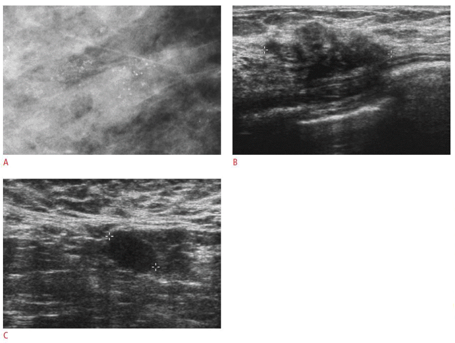
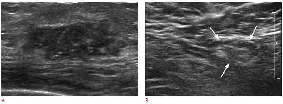
No differences were found between the underestimated and non-underestimated groups in terms of age, clinical symptoms, and mammographic findings. In the analysis of ultrasonographic findings, the rate of presence of abnormal lymph node depicted on ultrasonography in underestimated group was higher than that in the non-underestimated group (26.3% [5/19 lesions] vs. 4.9% [2/14 lesions], P=0.016, respectively) (Figs. 1, 2). No statistically significant differences were identified between the underestimated and non-underestimated groups with respect to ultrasonographic findings such as shape, orientation, margin, boundary, echogenicity, or microcalcification within the mass.
An axillary lymph node dissection or a sentinel lymph node biopsy was performed in all 60 patients. After surgery, none of the 41 patients in the non-underestimated group had detectable axillary lymph node metastases, whereas metastatic axillary lymph nodes were present in 3 of 19 patients in the underestimated group (P=0.021 by Fisher exact test). Among the seven patients determined to have abnormal axillary lymph nodes on ultrasonography, two patients in the underestimated group were shown to have lymph node metastasis following surgery. One metastatic axillary lymph node was actually a lymph node that had been evaluated as benign by US. The sensitivity and positive predictive value (PPV) of the axillary US findings with the histopathologic correlation of lymph node metastasis in the study were 66.7% (2/3) and 28.6% (2/7), respectively.
Although a US-14G-CNB is a highly accurate and widely used method for the diagnosis of breast lesions, sampling errors can result in the histologic underestimation of lesions containing atypical ductal hyperplasia (ADH) or DCIS, as well as invasive carcinomas. For a lesion diagnosed as ADH on needle biopsy and DCIS at surgery, underestimation is important because it could lead to a positive margin following surgical resection. Lesions diagnosed as DCIS on needle biopsy and unsuspected invasive carcinoma at surgery result in delayed lymph node biopsies. Thus, patients may have to undergo two separate surgical procedures: one procedure for excision of the lesion and an additional procedure for axillary lymph node evaluation [16].
It is useful to identify factors that can predict the DCIS underestimation after a US-14G-CNB to make more accurate surgical plans and to reduce the potential patient risk and overall medical costs. Variable DCIS underestimation rates ranging from 5%-44% and ADH underestimation rates of 11%-75% have been reported, and most biopsies have been performed using stereotactic devices with directional vacuum-assisted biopsy in addition to automated large-core needle biopsy [5-8,17]. However, DCIS underestimation by US-14G-CNB have not previously been sufficiently evaluated. In a review of the literature (Table 4), we identified the rates of DCIS underestimation in 10 studies following US-guided-CNB that ranged from 20% to 66.7%; the original aim of these studies was to evaluate the accuracy of a US-guided-CNB and not to determine the rate of DCIS underestimation [3,18-26]. In the present study, the rate of DCIS underestimation was 30.4% (21 of 69 lesions), which is within the range of previously published results. That higher rates of DCIS underestimation determined with the use of ultrasonography guidance were seen as compared with stereotactic biopsy techniques may be due to the fact that most US-guided biopsy procedures are performed on a mass, while, the most common indication for the use of a stereotactic biopsy is a microcalcification. The underestimation of invasive cancer is more frequent for a mass than for a microcalcification [17,27]. Some investigators have reported that 90% of carcinomas that present as microcalcifications alone were non-infiltrating, whereas 84% of carcinomas that present as a mass were invasive [28-30]. These investigators concluded that the presence of a mass lesion was a significant predictor for the presence of invasion.
It would be useful to identify the preoperative factors involved in predicting the presence of occult invasion within DCIS lesions. The ability to preoperatively identify patients with a high possibility of a co-existing invasive carcinoma might allow sentinel lymph node mapping and needle aspiration or a biopsy to be performed prior to the initial surgical excision. To identify possible factors involved in DCIS underestimation, we compared the underestimated and non-underestimated groups and found no statistically significant differences between the groups with regard to clinical and imaging characteristics, with the exception of the presence of abnormal axillary lymph nodes assessed by ultrasonography. Five of 19 patients (26.3%) in the underestimated group and two of 41 patients (4.9%) in the non-underestimated group demonstrated abnormal lymph nodes by axillary ultrasonography (P=0.016).
The accuracy of preoperative axillary ultrasonography for nodal metastasis in patients with invasive breast cancer has been reported in several studies with sensitivities ranging from 35% to 95% and PPVs ranging from 69% to 94.9%; these values are dependent on the association of specific diagnostic criteria, including lymph node shape, length and width, the appearance of the cortex and hilum, and the use of color Doppler ultrasonography to define suspicious lymph nodes on ultrasonography [31-33]. The sensitivity in our current study (66.7%) is within the previously reported ranges. Interestingly, the PPV in this study (28.6%) is lower than in previously published reports, which seems to be due to differences in the two study populations. While our study was confined to pure DCIS with the rare possibility of axillary lymph node metastasis, investigators in previous studies evaluated the axillae in patients with invasive breast cancers or extensive DCIS (at least 4 cm in extent), which are populations that already have a high likelihood of axillary lymph node metastases. A comparative analysis determining the usefulness of ultrasonographic criteria for predicting axillary nodal metastasis in abnormal lymph nodes in different settings with a high likelihood of an axillary metastasis or a low risk of lymph node metastasis is needed. DCIS is not an invasive malignancy and is not able to metastasize to the regional lymph nodes, and less than 1% to 2% of patients with DCIS who have undergone axillary lymph node dissection have been reported to have axillary lymph node metastasis [34,35]. In the current study, none of the 41 patients in the non-underestimated group who had undergone either axillary lymph node dissection or a sentinel lymph node biopsy had lymph node metastasis.
A multicenter study that examined factors related to DCIS underestimation after a stereotactic biopsy demonstrated that DCIS underestimation was more frequent in mass lesions as compared to lesions with microcalcifications (24.3% vs. 12.5%, respectively) [17]. In addition, they detected DCIS underestimation more frequently with the use of automated large-core devices than with the use of vacuum-assisted devices (20.4% vs. 11.2%, respectively), with ≤10 specimens as compared with >10 specimens (17.5% vs. 11.5%, respectively) and in lesions >20 mm as compared to lesions <10 mm(21.9% vs. 11.9%, respectively) [17]. Histologic factors, including high nuclear grade DCIS, comedo subtype, and large size have also been reported to significantly increase the likelihood of invasion found after surgical resection [27,36]. Recently, Park et al. [37,38] reported that underestimation was significantly related to lesion palpability, mass or calcification on ultrasonography, and core needle biopsy rather than vacuum-assisted biopsy and suggested using nomograms for predicting DCIS underestimation. A previous study by Lee et al. [9] reported that ultrasonographic lesions >20 mm in size were associated with invasive components on final pathology. However, in our study, the mean lesion size in the underestimated group (2.3 cm) was larger than in the non-underestimated group (1.6 cm), but this difference was not statistically significant (P=0.151).
This study had some limitations. First, the sample size of pure DCIS after a US-14G-CNB was relatively small. A further study with a greater number of cases diagnosed with DCIS after a US-14G-CNB is required. Second, the clinicians who performed the biopsies had varying levels of experience with the technique, which potentially could affect the underestimation rate. Third, the investigators were not blind to the biopsy results of the lesions being identified as pure DCIS, which could have influenced the retrospective review of the images.
In conclusion, the rate of DCIS underestimation in breast masses diagnosed as DCIS by a US-14G-CNB in this study was 30.4%. Our results underscore the difficulties in predicting possible pathologic underestimation solely relying on clinical and imaging findings of breast lesions. However, the presence of abnormal axillary lymph nodes on ultrasonography may be useful for preoperatively predicting DCIS underestimation.
Reference1. Parker SH, Jobe WE, Dennis MA, Stavros AT, Johnson KK, Yakes WF, et al. US-guided automated large-core breast biopsy. Radiology 1993;187:507–511.
2. Liberman L, Feng TL, Dershaw DD, Morris EA, Abramson AF. USguided core breast biopsy: use and cost-effectiveness. Radiology 1998;208:717–723.
3. Smith DN, Rosenfield Darling ML, Meyer JE, Denison CM, Rose DI, Lester S, et al. The utility of ultrasonographically guided largecore needle biopsy: results from 500 consecutive breast biopsies. J Ultrasound Med 2001;20:43–49.
4. Jackman RJ, Nowels KW, Shepard MJ, Finkelstein SI, Marzoni FA Jr. Stereotaxic large-core needle biopsy of 450 nonpalpable breast lesions with surgical correlation in lesions with cancer or atypical hyperplasia. Radiology 1994;193:91–95.
5. Liberman L, Dershaw DD, Rosen PP, Giess CS, Cohen MA, Abramson AF, et al. Stereotaxic core biopsy of breast carcinoma: accuracy at predicting invasion. Radiology 1995;194:379–381.
6. Jackman RJ, Nowels KW, Rodriguez-Soto J, Marzoni FA Jr, Finkelstein SI, Shepard MJ. Stereotactic, automated, large-core needle biopsy of nonpalpable breast lesions: false-negative and histologic underestimation rates after long-term follow-up. Radiology 1999;210:799–805.
7. Lee CH, Carter D, Philpotts LE, Couce ME, Horvath LJ, Lange RC, et al. Ductal carcinoma in situ diagnosed with stereotactic core needle biopsy: can invasion be predicted? Radiology 2000;217:466–470.
8. Rutstein LA, Johnson RR, Poller WR, Dabbs D, Groblewski J, Rakitt T, et al. Predictors of residual invasive disease after core needle biopsy diagnosis of ductal carcinoma in situ. Breast J 2007;13:251–257.
9. Lee JW, Han W, Ko E, Cho J, Kim EK, Jung SY, et al. Sonographic lesion size of ductal carcinoma in situ as a preoperative predictor for the presence of an invasive focus. J Surg Oncol 2008;98:15–20.
10. Silver SA, Tavassoli FA. Mammary ductal carcinoma in situ with microinvasion. Cancer 1998;82:2382–2390.
11. American College of Radiology. Breast Imaging Reporting and Data System® (BI-RADS®) Atlas. 4th ed. Reston, VA: American College of Radiology, 2003.
12. Feu J, Tresserra F, Fabregas R, Navarro B, Grases PJ, Suris JC, et al. Metastatic breast carcinoma in axillary lymph nodes: in vitro US detection. Radiology 1997;205:831–835.
13. Vassallo P, Wernecke K, Roos N, Peters PE. Differentiation of benign from malignant superficial lymphadenopathy: the role of highresolution US. Radiology 1992;183:215–220.
14. Ecanow JS, Abe H, Newstead GM, Ecanow DB, Jeske JM. Axillary staging of breast cancer: what the radiologist should know. Radiographics 2013;33:1589–1612.
15. Berry CC. A tutorial on confidence intervals for proportions in diagnostic radiology. AJR Am J Roentgenol 1990;154:477–480.
16. Liberman L, Goodstine SL, Dershaw DD, Morris EA, LaTrenta LR, Abramson AF, et al. One operation after percutaneous diagnosis of nonpalpable breast cancer: frequency and associated factors. AJR Am J Roentgenol 2002;178:673–679.
17. Jackman RJ, Burbank F, Parker SH, Evans WP 3rd, Lechner MC, Richardson TR, et al. Stereotactic breast biopsy of nonpalpable lesions: determinants of ductal carcinoma in situ underestimation rates. Radiology 2001;218:497–502.
18. Schueller G, Jaromi S, Ponhold L, Fuchsjaeger M, Rudas M, et al. US-guided 14-gauge core-needle breast biopsy: results of a validation study in 1352 cases. Radiology 2008;248:406–413.
19. Cho N, Moon WK, Cha JH, Kim SM, Kim SJ, Lee SH, et al. Sonographically guided core biopsy of the breast: comparison of 14-gauge automated gun and 11-gauge directional vacuumassisted biopsy methods. Korean J Radiol 2005;6:102–109.
20. Sauer G, Deissler H, Strunz K, Helms G, Remmel E, Koretz K, et al. Ultrasound-guided large-core needle biopsies of breast lesions: analysis of 962 cases to determine the number of samples for reliable tumour classification. Br J Cancer 2005;92:231–235.
21. Crystal P, Koretz M, Shcharynsky S, Makarov V, Strano S. Accuracy of sonographically guided 14-gauge core-needle biopsy: results of 715 consecutive breast biopsies with at least two-year follow-up of benign lesions. J Clin Ultrasound 2005;33:47–52.
22. Pijnappel RM, van den Donk M, Holland R, Mali WP, Peterse JL, Hendriks JH, et al. Diagnostic accuracy for different strategies of image-guided breast intervention in cases of nonpalpable breast lesions. Br J Cancer 2004;90:595–600.
23. Philpotts LE, Hooley RJ, Lee CH. Comparison of automated versus vacuum-assisted biopsy methods for sonographically guided core biopsy of the breast. AJR Am J Roentgenol 2003;180:347–351.
24. Schoonjans JM, Brem RF. Fourteen-gauge ultrasonographically guided large-core needle biopsy of breast masses. J Ultrasound Med 2001;20:967–972.
25. Buchberger W, Niehoff A, Obrist P, Rettl G, Dunser M. Sonographically guided core needle biopsy of the breast: technique, accuracy and indications. Radiologe 2002;42:25–32.
26. Youk JH, Kim EK, Kim MJ, Oh KK. Sonographically guided 14-gauge core needle biopsy of breast masses: a review of 2,420 cases with long-term follow-up. AJR Am J Roentgenol 2008;190:202–207.
27. King TA, Farr GH Jr, Cederbom GJ, Smetherman DH, Bolton JS, Stolier AJ, et al. A mass on breast imaging predicts coexisting invasive carcinoma in patients with a core biopsy diagnosis of ductal carcinoma in situ. Am Surg 2001;67:907–912.
28. Sener SF, Candela FC, Paige ML, Bernstein JR, Winchester DP. Limitations of mammography in the identification of noninfiltrating carcinoma of the breast. Surg Gynecol Obstet 1988;167:135–140.
29. Fuhrman GM, Cederbom GJ, Bolton JS, King TA, Duncan JL, Champaign JL, et al. Image-guided core-needle breast biopsy is an accurate technique to evaluate patients with nonpalpable imaging abnormalities. Ann Surg 1998;227:932–939.
30. Dillon MF, McDermott EW, Quinn CM, O'Doherty A, O'Higgins N, Hill AD. Predictors of invasive disease in breast cancer when core biopsy demonstrates DCIS only. J Surg Oncol 2006;93:559–563.
31. Yang WT, Ahuja A, Tang A, Suen M, King W, Metreweli C. High resolution sonographic detection of axillary lymph node metastases in breast cancer. J Ultrasound Med 1996;15:241–246.
32. Bedrosian I, Bedi D, Kuerer HM, Fornage BD, Harker L, Ross MI, et al. Impact of clinicopathological factors on sensitivity of axillary ultrasonography in the detection of axillary nodal metastases in patients with breast cancer. Ann Surg Oncol 2003;10:1025–1030.
33. Abe H, Schmidt RA, Kulkarni K, Sennett CA, Mueller JS, Newstead GM. Axillary lymph nodes suspicious for breast cancer metastasis: sampling with US-guided 14-gauge core-needle biopsy: clinical experience in 100 patients. Radiology 2009;250:41–49.
34. Silverstein MJ, Skinner KA, Lomis TJ. Predicting axillary nodal positivity in 2282 patients with breast carcinoma. World J Surg 2001;25:767–772.
35. Zelis JJ, Sickle-Santanello BJ, Liang WC, Nims TA. Do not contemplate invasive surgery for ductal carcinoma in situ. Am J Surg 2002;184:348–349.
36. Patchefsky AS, Schwartz GF, Finkelstein SD, Prestipino A, Sohn SE, Singer JS, et al. Heterogeneity of intraductal carcinoma of the breast. Cancer 1989;63:731–741.
A 42-year-old woman with a palpable mass in her right breast which proved to be an invasive ductal carcinoma.A. Mammogram demonstrates suspicious microcalcifications in the right breast. B. Breast ultrasonography reveals a 3 cm, irregularly-shaped and microlobulated margined hypoechoic mass with echogenic foci within the right upper outer quadrant. C. Abnormal lymph nodes without fatty hilum in the right axilla can be seen. The patient underwent an ultrasound-guided 14-gauge core needle biopsy with a ductal carcinoma in situ identified based on the subsequent histology. Invasive carcinoma was found following surgical excision with the presence of metastatic lymph nodes detected after axillary lymph node dissection.
 Fig. 1.A 42-year-old woman with ductal carcinoma in situ.A, B. Ultrasonograms demonstrate an irregularly-shaped, hypoechoic mass in the right upper outer portion with a normal appearing lymph node (arrows) in the right axilla. The pathologic findings following an ultrasound-guided 14-gauge core needle biopsy and surgical excision were consistent with ductal carcinoma in in situ.
 Fig. 2.Table 1.Comparisons of clinical findings in the 21 underestimated and 48 non-underestimated DCIS lesions Table 2.Comparisons of mammographic findings in the underestimated and non-underestimated groups Table 3.Comparisons of ultrasonographic findings in the underestimated and non-underestimated groups Table 4.Reported rates of DCIS underestimation after an ultrasound-guided 14-gauge core needle biopsy
|


 Print
Print facebook
 twitter
 Linkedin
 google+
 Download Citation
 PDF Links
 PubReader
 ePub Link
 Full text via DOI
 Full text via PMC


