

 Print
Print facebook
 twitter
 Linkedin
 google+

Introduction
Primary thyroid lymphoma (PTL) is a relatively uncommon pathology of the thyroid gland, with generally good prognosis if treated appropriately. There have been several reports on the imaging characteristics of PTL: homogeneously hypoechoic mass with posterior enhancement on ultrasonography, and homogenously enhancing mass without invasion to the surrounding structures on computed tomography (CT) scan, hardly accompanying intratumoral calcification, cystic degeneration, or necrosis [1-5]. However, sometimes PTL may show aggressive natures such as rapid growth and invasion of adjacent structures. To our knowledge, there are only a few literatures about image features of aggressive PTL [1]. Here we describe image features of two cases of aggressive PTL which mimicked image features of anaplastic thyroid cancer (ATC).
Case Reports
Case 1
A 77-year-old woman presented to our outpatient clinic with a two-month history of dysphasia. On physical examination, both thyroid lobes were diffusely enlarged with the right lobe being slightly firmer than the left lobe. There were palpable masses along the right cervical levels II and III. Thyroid function test was within normal range. She had no medical history of Hashimoto thyroiditis. Flexible fiberoptic laryngoscopy showed no remarkable finding. We supposed thyroid cancer with lymph node metastasis.
On ultrasonography (iU-22, Philips Medical System, Bothwell, WA, USA) with high frequency linear-array transducer, a larger than 4-cm, irregular shaped, heterogeneously hypoechoic, solid mass occupying the entire right thyroid lobe was seen with suspicious several small internal calcifications. The mass directly invaded the right lateral and posterior wall of trachea and encased right common carotid artery (Fig. 1A, B). We presupposed anaplastic carcinoma, and ultrasoundguided fine needle aspiration (FNA) was carefully performed. The cytologic results revealed suspicious finding for undifferentiated carcinoma. Contrast-enhanced CT of the neck showed a heterogeneously enhancing mass compared to adjacent muscle in the posterior portion of the right thyroid lobe, outgrowing toward the posterior wall of the trachea. There was no grossly defined calcification, hemorrhage, or nonenhancing necrotic portion within the mass. The mass showed indistinct margins with the proximal esophagus (Fig. 1C). Magnetic resonance imaging (MRI) of the neck was done to evaluate esophageal and tracheal invasion. The mass showed heterogeneous enhancement with focal low signal intensity portion, suggesting suspicious necrotic portion. The tumor invaded the proximal esophagus, extended anteriorly to the strap muscle, and encased right common carotid artery (Fig. 1D). These findings suggested ATC with tracheal and vascular invasion with suspicious esophageal invasion. A few enhancing small lymph nodes were seen at the left level II and III with the short diameter of 0.6 cm. Subsequent excisional biopsy of the thyroid lesion was done, and it was histopathologically diagnosed as diffuse large B cell lymphoma.
The patient received three cycles of R-CHOP (rituximab, cyclophosphamide, adriamycin, vincristine, and prednisolone) chemotherapy as well as total 3,816 cGy of intensity-modulated radiation therapy (IMRT), which resulted in complete regression on the CT images after 4 months from the initial diagnosis.
Case 2
A 73-year-old woman was transferred to our institution with persisting voice change for eight months. Flexible fiberoptic laryngoscopy performed at our institution revealed intraluminal mass emerging from the left posterolateral aspect of the subglottis, cricoid cartilage, and trachea, implying suspicious invasion by left paratracheal mass. Left vocal cord palsy was also noted.
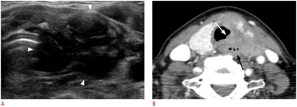
Ultrasonography of neck showed a larger than 4-cm, irregular shaped, heterogeneous hypoechoic mass in the left thyroid gland. The mass extended to the left posterolateral wall of trachea and the left extrathyroid space (Fig. 2A). Thyroid function test was within normal range. There was no medical history of Hashimoto thyroiditis. We supposed ATC, and ultrasound-guided FNA was carried out for the left thyroid mass. The cytology suggested suspicious for malignancy.
CT scan demonstrated an irregular shaped, isodense mass involving the left lobe and isthmus of the thyroid gland, without internal calcification or hemorrhage. Extrathyroid extension was present multi-directionally with adjacent organ invasion; strap muscles anteriorly, tracheal lumen medially, closely abutting the esophagus posteriorly which raised the possibility of esophageal invasion, and left pyriform sinus superiorly. There was no abnormal cervical lymph node (Fig. 2B). MRI of the neck was additionally performed, allowing more distinct demarcation of the lesion boundaries of the known thyroid mass with tracheal, laryngeal, and esophageal invasion. The mass showed heterogeneous enhancement and extend to the lateral and posterior direction. The esophagus had been encroached on by the mass, with loss of the tissue plane between them and this finding allowed diagnosing esophageal invasion. The mass also closely abutted the left internal jugular vein and common carotid artery, but there was no definite vascular invasion. Incisional biopsy was done for the mass, and the lesion was pathologically confirmed as diffuse large B cell lymphoma.
The patient received four cycles of R-CHOP chemotherapy, followed by total 4,000 cGy of IMRT, which resulted in complete regression on the CT images after 4 months from the initial diagnosis.
Discussion
PTL is relatively rare, representing less than 5% of thyroid malignancies. Common clinical feature of thyroid lymphoma is rapidly growing mass-like anaplastic carcinoma in middle-aged to elderly women, especially when a history of long-standing thyroiditis is present. Although PTL may grow suddenly and be life-threatening due to airway obstruction, early detection and diagnosis of it followed by appropriate treatment can lead to favorable prognosis [6].
The typical ultrasonographic feature of PTL is diffuse hypoechoic parenchyma with intervening echogenic septa-like structures or circumscribed mass, which is usually limited to one lobe with markedly hypoechoic and homogenous echogenicity. Posterior acoustic enhancement is another distinguishing feature of thyroid lymphoma, and cervical or mediastinal lymphadenopathies are accompanied in many cases [1,2,4,5]. Invasion to the surrounding structures is rare. In the ultrasonography of our first case, some microcalcifications were suspected to be present in the mass. However, no calcification was detected in both subsequent CT and pathologic examination. Such ultrasonographic finding seemed to be a pseudolesion caused by heterogenous echogenicity of tumor mass.
The primary differential diagnosis for thyroid lymphoma presenting as a rapidly growing large mass in elderly persons is ATC, which comprises only 1%-2% of thyroid malignancies and shows extremely poor prognosis with a mean survival of 6 months [7-9]. As with thyroid lymphoma, ATC may show rapid progression, resulting in aerodigestive tract compromise. On imaging features, ATC is presented as a large, solid mass accompanied by necrosis, hemorrhage, dense calcification, direct invasion into adjacent organs, and cervical lymph node metastases. Tumor necrosis has been known to be the most valuable parameter in differentiating ATC from other thyroid masses, and low attenuation value on postcontrast scan (attenuation value<100 HU) is another predictor of anaplastic carcinoma [7-10].
Although, ultrasonography is the modality of choice for imaging of the thyroid pathology, it is less effective to evaluate large thyroid masses. Ultrasonographic features of our cases are heterogeneous and hypoechoic mass with extensive invasion to the adjacent structures; these findings imply ATC rather than lymphoma. Previously reported ultrasonographic features of PTL are homogenous hypoechoic mass with rare invasion of the adjacent structures [2,4,5]. These findings were quite different from our cases. Moreover, our cases demonstrated heterogeneous enhancement of the masses, which was discordant with the well-known homogeneous appearance of lymphoma [2,3]. MRI did not help much in differentiating PTL from ATC in our cases, as MRI of our cases also showed heterogeneous enhancement and some suspicious necrotic portion in the mass. These findings indicate that imaging findings alone are not sufficient to make distinction between PTL and ATC, when the lesion displays aggressive appearance at the time of diagnosis.
Ultrasound-guided FNA is the first step in diagnostic strategy for a thyroid nodule, but the accuracy is quite low when diagnosing thyroid lymphoma or anaplastic cancer. Core needle biopsy (CNB), which provides enough tissue needed for the exact diagnosis of aggressive thyroid cancer such as lymphoma or anaplastic cancer and makes it possible to perform additional assays, could be the best primary approach for diagnosis of thyroid lymphoma compared with the FNA. Therefore, CNB should be the first step of diagnostic approach when a large aggressive thyroid mass is encountered on ultrasonography and CT evaluation [5,6,9].
In conclusion, we described image features of two cases of aggressive PTL in elderly patients, exhibiting heterogeneous echogenicity with extensive invasion to the adjacent structures. In addition, the masses showed heterogeneous attenuation on postcontrast CT scan. The ultrasonographic features of aggressive PTL were not specific enough to differentiate PTL from other type of aggressive thyroid cancer such as ATC in the elderly patients. Thus, more reliable first line diagnostic approach such as ultrasoundguided CNB should be performed as well as image studies to make accurate diagnosis.

 Download Citation
 PDF Links
 PubReader
 ePub Link
 Full text via DOI
 Full text via PMC


