

 Print
Print facebook
 twitter
 Linkedin
 google+

Introduction
When color Doppler images are obtained for static scattering targets, twinkling colors may appear due to the presence of turbulent flows [1,2]. This effect is often referred to as the presence of twinkling artifacts. The mechanisms underlying color Doppler twinkling artifacts are not clearly understood yet, even though the twinkling is known to be target-dependent and is caused by noises that arise from ultrasonic scanners [3]. Recently, the acoustic radiation force (ARF) has been proposed as the potential source of color Doppler twinkling artifacts, but this has not yet been experimentally verified [4].
The ultrasound used to construct color Doppler images produces an ARF that pushes targets, which are also known as scattering particles. The resulting target movement induces a frequency shift known as the Doppler shift in the ultrasound that is received. In such a case, the target movement resulting from the ARF is repeated at the frequency of a given pulse repetition frequency (PRF). This implies that Doppler shifts resulting from the ARF are narrowbanded and are centered at the PRF.
Users in the color Doppler mode cannot readily control the ARF, making it difficult to clinically examine the effects of radiation force on twinkling artifacts. However, the PRF, which reflects the frequency of the ARF that is exerted on targets, can be set by users. Careful observation of twinkling colors in relation to the PRF may provide insights as to whether color Doppler twinkling artifacts are associated with the ARF. This study aimed to experimentally investigate the significance of the ARF as a potential source of twinkling artifacts in color Doppler images.
Materials and Methods
Twinkling images were obtained at varying PRFs on a highcontrast circular static target containing highly echogenic scattering particles. In order to examine the influence of the PRF on the ARF, the transmitting ultrasounds produced by the ultrasonic scanner were measured in a water tank using a hydrophone. Based on these experimental results, the role of the ARF on color Doppler twinkling artifacts was assessed.
Color Doppler Imaging of a Scattering Contrast Target
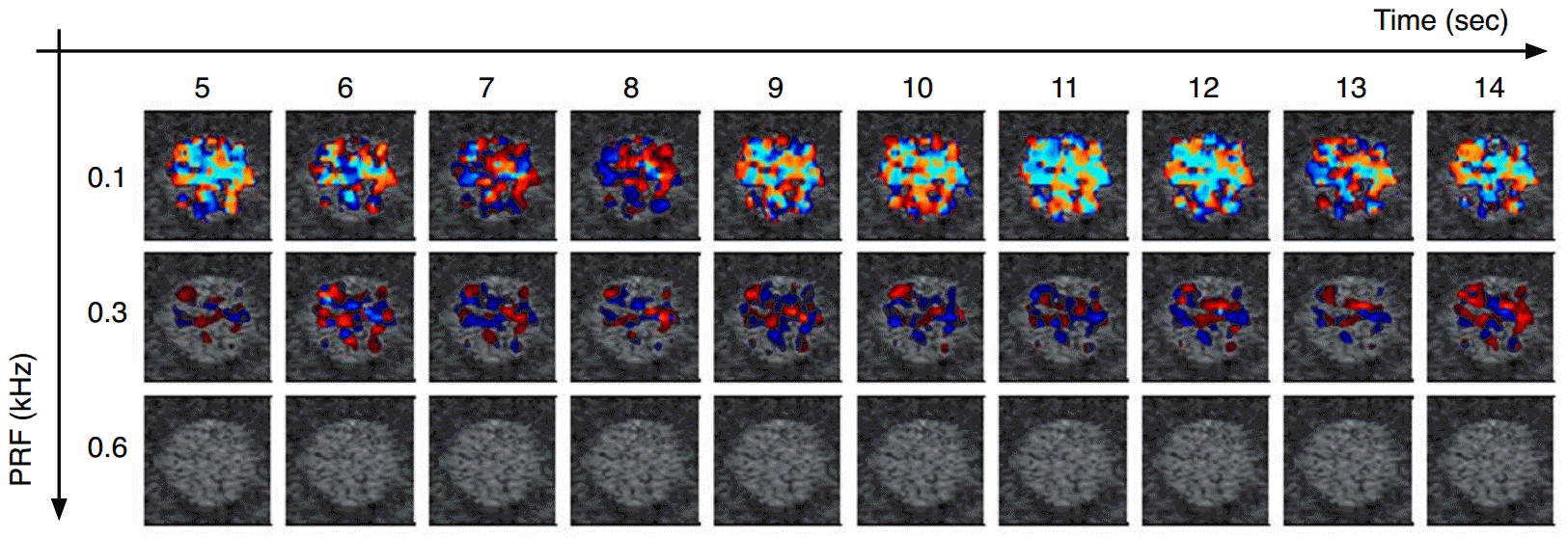
A color Doppler image was obtained of a circular scattering target using a GE ultrasound scanner (Voluson e, GE Healthcare, Oberösterreich, Austria) with a linear probe (12L-RS, 3-12 MHz). The scattering target was taken from the +15 dB circular contrast target of a commercial ultrasound standard quality assurance phantom (Model 551 small parts phantom, ATS Laboratories Inc., Bridgeport, CT, USA). The diameter of the target was 6 mm. The phantom was placed on a vibration-free supporter (RFB, NTR Systems, Seattle, WA, USA) to isolate the phantom from external vibrations. Ultrasonic coupling gel was applied to the upper surface of the phantom before scanning. The focal zone of the probe was set at the center of the target [5-8]. Color Doppler images were acquired under the default settings of the control parameters except for the PRF (e.g., Doppler frequency low, Doppler gain 0, and wall motion filter high). The extent of the twinkling was quantified by the number of color pixels within the target [9,10]. The color pixel number was counted using MatLab (MATLAB R2009a, MathWorks Inc., Natick, MA, USA).
Measurements of Transmitted Ultrasonic Pulses
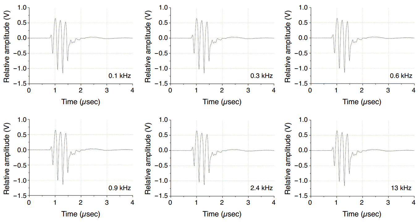
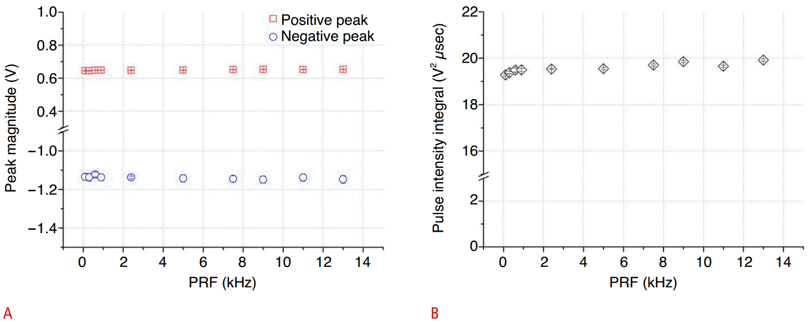
The ARF that can induce a target to vibrate at the PRF is produced by each transmitted ultrasonic pulse that irradiates a target when the ultrasonic scanner is operated in the color Doppler mode. The ARF is estimated from the transmitted ultrasonic pulse and the acoustic properties of the medium through which the ultrasound pulse propagates. For a given imaging target, the ARF is approximately derived from the transmitted ultrasonic pulse measured in a reference medium. The transmitted pulse was measured in a water tank using a needle hydrophone (TNU001A, NTR Systems) at nominal PRFs ranging from 0.1 kHz to higher values (Fig. 1). The measured transmitted pulses were stored on a digital scope (CS122G1, GaGe, Dynamic Signals LLC, Lockport, IL, USA). The pulse intensity integral of each transmitting ultrasonic pulse was calculated from the measured waveform, which is closely related to the ARF [11]. The recordings were repeated 12 times in order to examine the variability of the pressure and the intensity of transmitted ultrasonic pulses.
Results
Typical transmitted ultrasonic pulses produced by the clinical ultrasonic scanner operated in color Doppler mode are shown in Fig. 2, measured in a water tank with nominal PRFs set to 0.1 kHz, 0.3 kHz, 0.6 kHz, 0.9 kHz, 2.4 kHz, and 13 kHz. These ultrasonic pulses lasted for three to four cycles at a center frequency of approximately 5 MHz and had negative peak pressures about twice the corresponding positive peak pressures. The measured waveforms, regardless of the PRF, were found to be almost identical.
As expected, the mean values of the peak pressures and the pulse intensity integral remained virtually unchanged (within 3%) as the PRF varied from 0.1 to 13 kHz (Fig. 3). The standard deviations were found to be insignificant, indicating that the transmitted ultrasonic pulses produced for color Doppler imaging were highly stable. The PRF was not observed to have an effect on transmitted ultrasonic pulses, which suggests that the PRF also does not affect the magnitude of the ARF produced by the transmitted ultrasonic pulse that irradiates the target.
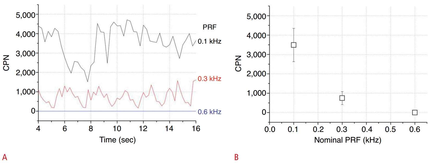
Typical color Doppler images (Fig. 4) were obtained for the circular +15 dB scattering contrast target at the nominal PRF values of 0.1, 0.3, and 0.6 kHz, over for a period extending up to 14 seconds from the onset of image acquisition. Less twinkling appeared as the PRF increased and twinkling artifacts could not be seen at PRF settings of 0.6 kHz or higher in color Doppler images (Fig. 4). The twinkling colors changed randomly over time, which can be attributed to random Doppler noises. This change is illustrated by the time history of the color pixel number within the target, which was displayed for 12 seconds at the nominal PRF settings of 0.1, 0.3, and 0.6 kHz (Fig. 5A). The temporal mean values of the extent of color twinkling were highest at the lowest PRF (0.1 kHz) and exponentially decreased as the PRF increased. A similar trend was found in the temporal variability of the twinkling plotted against the PRF (Fig. 5B).
Discussion
The twinkling artifact in color Doppler images can be easily seen at the rear of strong reflective targets, including renal stones [5,6,12-14] and guided needle punctures [15], as well as around or inside scattering soft tissue lesions such as calcified pancreatitis [15] and liver hyperechoic masses [15,16]. This suggests that color Doppler twinkling artifacts are associated with the echogenicity of targets and the acoustic impedance mismatch between the targets and the background. The impedance mismatch may cause a significant ultrasonic wave reflection at the interface of the target, possibly resulting in a radiation force directed toward the target.
If it were the case that a transmitted ultrasonic pulse produces the ARF exerted on a target, causing it to vibrate, the resulting alternating Doppler shifts displayed on the image as color twinkling would be narrow-banded. The spectra of the Doppler signals would be centered at the PRF, because the ARF is repeatedly exerted on the target to induce vibration at the PRF. The narrow band, however, is located far beyond the range of the color scale bar (from -PRF/2 to +PRF/2), and thus results in color (spectral) aliasing. The aliasing is displayed on the image with twinkling colors. If the ARF induces the aliasing, the resulting twinkling color would be expected to appear at all values of the PRF. In the present study, however, the twinkling artifacts appeared when the PRF was either 0.1 or 0.3 kHz, but completely disappeared when the PRF exceeded 0.6 kHz (Figs. 4, 5). This corresponds to previous observations that color Doppler twinkling artifacts highly dependent on the PRF of the ultrasound transmission, as reported by Kamaya et al. [10].
It should be noted that contradictory findings have been reported regarding the relationship between twinkling artifacts and the PRF. One group of researchers [17,18] has reported that more twinkling appeared at higher PRFs, whereas other investigators [3,7,9] have claimed the opposite results. Such contradictory observations can probably be attributed to differences in the targets and ultrasonic scanners used in each study.
In the present study, the measured transmitted ultrasonic pulses remained virtually unchanged while the PRF varied from 0.1 to 13 kHz (Figs. 2, 3). This suggests that the PRF does not affect the magnitude of the ARF induced by the transmitted ultrasonic pulse that irradiates the target, as the ARF is proportional to the pulse intensity integral. Linking these two observations (first, that the ARF was insensitive to the PRF and second, that the extent of color Doppler twinkling was highly affected to the PRF) leads to the deduction that the ARF is unlikely to be associated with color Doppler twinkling artifacts. This deduction is valid for the present experimental conditions performed on a static scattering contrast target with a clinical ultrasonic scanner. Further verification is required on other clinical scanners and in other conditions.
In summary, the color Doppler twinkling that appeared in a circular scattering target was observed to depend on the PRF setting. The PRF, however, was found to be insensitive to the ARF resulting from each transmitted ultrasonic pulse that irradiated the imaging target. No evidence was found of ARF-induced twinkling aliasing, which, if occurred, was expected to appear at all values of PRF. These results suggest that the ARF is unlikely to be associated with color Doppler twinkling artifacts, and therefore, that the ARF cannot be considered as a potential source for color twinkling artifacts. This conclusion is valid under the present experimental conditions for color Doppler imaging, and further verification is required with regard to other clinical conditions.

 Download Citation
 PDF Links
 PubReader
 ePub Link
 Full text via DOI
 Full text via PMC


