

 Print
Print facebook
 twitter
 Linkedin
 google+

Introduction
Ultrasound (US) elastography refers to a noninvasive imaging technique that can describe tissue displacement (i.e., strain) or stiffness in response to a given force [1,2]. Stiff tissues tend to deform less and show less strain than compliant tissues in response to the same imparted force. Therefore, the fundamentals of US elastography are similar to manual palpation [2]. Since its introduction, US elastography has enabled the evaluation of many different types of organs, including the breast, liver, prostate, thyroid glands, blood vessels, salivary glands, musculoskeletal structures, and cervical lymph nodes. At the time of writing, more than 10 pilot studies have been published on the use of US elastography to evaluate the cervical lymph nodes. This review details the currently available techniques for assessing the cervical lymph nodes and describes the main published results, reproducibility, and limitations of US elastography in clinical practice.
US Elastography Techniques
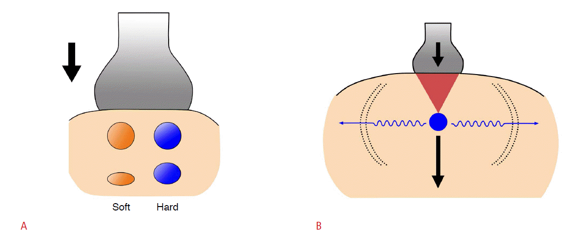
US elastography techniques can be broadly divided into two groups: strain elastography and shear wave-based elastography. Strain elastography measures tissue displacement along the axis of the applied force. This type of displacement can be found in an elastic spring that is compressed or stretched along its axis. Shear wave-based elastography is a recently developed elastography technique and measures a different type of wave that is generated when tissues are mechanically stimulated by focused pulses, though shear waves are relatively slow and weak waves that are produced by the tangential propagation of the tissue and move perpendicular to the direction of the imparted force (Fig. 1) [3].
Strain Elastography
Strain elastography was the first commercialized elastography technique to be developed to assess stiffness noninvasively and has received considerable attention because it can be performed using conventional US hardware with software modifications. Axial tissue displacement can be caused by either freehand compression along the axis of the US beam or stationary and endogenous stresses such as cardiovascular movements or other physiologic motions [4]. Strain elastography requires calculating tissue displacement along the beam axis in response to an applied force. Dedicated software is required to obtain the comparative difference in tissue movement from one frame to another, and then measure the tissue strain, which can be expressed as change in length divided by the original length. The resulting strain data are graphically displayed as a two-dimensional map of the relative tissue strain, which is called an elastogram. Elastograms are usually displayed as a semitransparent overlay onto gray-scale images using a color-coded scale that can vary among US systems [3,5].
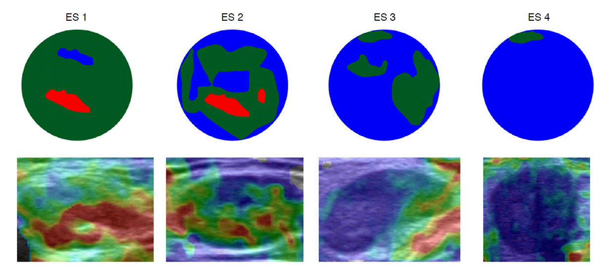
Using strain elastography, two kinds of elasticity assessments can be obtained. First, visual scoring of the colors within and around the lesions can be assessed using a 4-5-point elastographic scale, which measures the relative proportion of the intralesional lowstrain areas (i.e., high stiffness); therefore, the stiffer the tissue is, the higher the elastographic scale scores assigned. Using this system, elastographic scale 1 refers to a soft nodule that is predominantly red, with <10% of the area colored as blue; elastographic scale 2 refers to a moderately soft nodule that is predominantly red and green, with 10%-50% of the area shown as blue; elastographic scale 3 refers to a moderately stiff nodule that is predominantly blue and green, with 50%-90% of the area shown as blue; and elastographic scale 4 refers to a stiff nodule that is predominantly (>90%) blue (Fig. 2) [6-11]. Depending on the specific US machine, these color scales may be inversely applied. In a second method, two regions of interest (ROIs) are drawn over the target region and an adjacent reference region consisting of normal muscle or subcutaneous fat. The strain ratio is then automatically computed by the US machine (Fig. 3). Strain ratios are normally calculated such that values >1 indicate that the target lesion has a lower strain than the reference tissue, indicating higher stiffness. The likelihood of malignancy increases as the strain ratio increases [7,8].
Shear Wave-Based Elastography
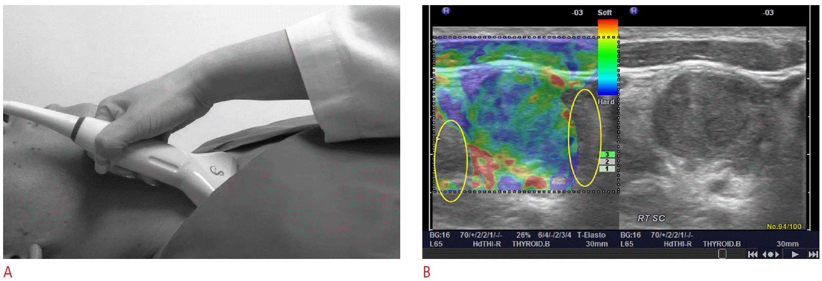
Unlike the use of freehand compression in strain elastography, in shear wave-based elastography, focused high-intensity, short-duration acoustic pulses from a US transducer make shear waves by absorbing acoustic energy. In order to obtain the appropriate images, the probe is applied with the minimum amount of pressure required to make complete contact with the cervical lymph node. The velocities of the shear waves are tracked by a series of normal-intensity impulses using US correlation methods. According to the following formula, it is inferred that the velocities of the shear waves are determined by the tissue stiffness: E=3pc2, where E is stiffness (i.e., Young’s elastic modulus [kPa]), c is the shear wave speed (m/sec), and p is the density (kg/m3). The density of most soft tissues is about the same as that of water (1,000 kg/m3).
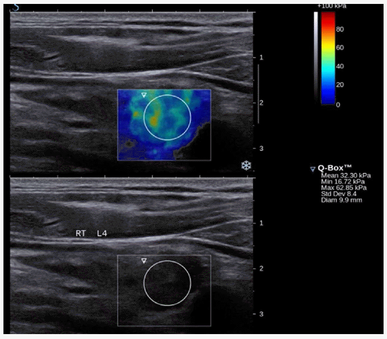
The precise implementation of shear wave-based elastography technology differs among US system manufacturers and involves various techniques, such as acoustic radiation force impulse imaging and supersonic shear imaging. Acoustic radiation force impulse imaging generates shear waves with a single pushing beam. This system does not currently display real-time elastograms. The quantitative shear wave speed (m/sec) can be determined in ROIs that are typically 5×6 mm boxes, which are relatively small compared to those used in supersonic shear imaging, and displayed within static elastograms (Fig. 4) [12-15]. Supersonic shear imaging generates supersonic speed pulses that induce and amplify shear waves, which are then tracked throughout the entire window using ultrafast US tracking impulses and computationally demanding post-processing software. This system displays real-time, color-coded elastograms of the shear wave speed (m/sec) or elastic modulus (kPa), and quantitative measurements can be obtained for ROIs using static elastograms (Fig. 5) [16].
Clinical Applications
Several pilot studies have evaluated the ability of US elastography to detect malignancies in the cervical lymph nodes, including a variety of benign and malignant pathologies [6,8,10,11,17-27]. However, there is marked selection bias in these studies since US elastography was deployed for pathologic verification or to assess enlarged or abnormal US findings, thereby resulting in samples that included a high proportion of malignancies (41%-85%). Table 1 summarizes the main characteristics of the previously published studies.
Lyshchik et al. [8] published one of the first clinical studies to evaluate the diagnostic promise of strain elastography by examining 141 lymph nodes, of which 98 were confirmed as benign and 43 were confirmed as malignant by histopathology. They classified the nodes according to visualization, brightness compared to the neighboring muscles, and the regularity of the outline. Using a strain ratio cut-off value of >1.5, strain elastography demonstrated sensitivity, specificity, and accuracy values of 85%, 98%, and 92%, respectively. According to a recent meta-analysis of nine strain elastography studies that included 50-155 cervical or axillary lymph nodes [7], the pooled sensitivity and specificity values for detecting malignancies were 74% (95% confidence interval [CI], 66% to 81%) and 90% (95% CI, 82% to 94%) for elastographic scale and 88% (95% CI, 79% to 93%) and 81% (95% CI, 49% to 95%) using strain ratios, respectively. The 4-point elastography scale is most frequently used for detecting malignant lymph nodes. In general, metastatic lymph nodes demonstrate higher stiffness than benign lymph nodes, so elastographic scale scores of 1-2 indicate benign lymph nodes, and elastographic scale scores of 3-4 indicate malignant lymph nodes [6]. To account for the possibility that intranodal necrosis in metastatic lymph nodes might confound elastographic scale, some investigators have modified their qualitative scoring systems to classify nodes that demonstrate a specific strain pattern (using 5-point scale), peripheral low strain (shown by high stiffness), or central high strain (shown by low stiffness). Ishibashi et al. [10] assessed elastographic scale using a 5-point scale and documented that elastography in combination with conventional US demonstrated sensitivity, specificity, and accuracy values of 90.3%, 80%, and 84.5%, respectively. Similarly, Alam et al. [11] used a 5-point scale for elastography and reported sensitivity, specificity, and accuracy values of 83%, 100%, and 89%, respectively.
The strain elastography technique can help differentiate reactive and metastatic lymph nodes, but cannot distinguish other conditions such as lymphoma or tuberculous lymphadenitis [7]. Lymphomas produce softer-appearing nodes [19], and tuberculous lymphadenitis with scarring calcification or necrosis may demonstrate stiffer areas or a mixture of colors [18]. Little is known about the strain elastographic appearance of relapsing or chronic lymphadenitis or rare benign diseases such as Kikuchi or Kimura disease. According to a recent study, Kikuchi disease demonstrates a soft appearance (i.e., elastographic scale 1) on strain elastography, which would suggest a benign lesion [28].
There are relatively few clinical studies that have compared acoustic radiation force impulse imaging and supersonic shear imaging [24-27,29]. Bhatia et al. [27] has reported that the median elastic modulus of malignant lymph nodes is higher than that of benign lymph nodes. However, discrimination was low because the optimal cut-off value of 30.2 kPa demonstrated sensitivity, specificity, and accuracy values of 41.9%, 100%, and 61.8%, respectively. Another study has reported that the maximum elastic modulus can be used to differentiate malignant lymph nodes, and that a cut-off value of 19.4 kPa resulted in accuracy, sensitivity, and specificity values of 94%, 91%, and 97%, respectively [26]. However, since only a few preliminary papers have been published on this topic, further research is required, although most studies do suggest that US elastography may be useful for diagnosing lymph nodes.
Reproducibility and Limitations
Strain elastography using freehand compression is very dependent on the compression technique, because excessive compression alters tissue stiffness and nonaxial displacement (i.e., displacement that occurs laterally or outside of the imaging plane) can degrade the accuracy of the software’s correlation algorithms (Fig. 6). Because shear wave-based elastography does not require freehand compression, it may be less operator-dependent than strain elastography [30]. Lenghel et al. [19], Tan et al. [20], and Bhatia et al. [21] have reported interobserver agreement values of 0.374-0.738, 0.828-0.946, and 0.687, respectively, for strain elastography (as indicated by weighted κ values), which suggest fair to almost perfect agreement. For shear wave-based elastography, intraobserver and interobserver reproducibility are moderate to excellent according to the calculated intraclass correlation coefficients (intraobserver, 0.64 to 0.84; interobserver, 0.72 to 0.77) [29]. Nevertheless, US elastography has several unresolved issues and limitations. First, US elastography can be problematic if there is a focal convex bulge on the skin overlying the ROI because, under these circumstances, it may be impossible to apply a linear transducer without producing focal stress concentrations within the tissue of interest, thereby resulting in spuriously stiff elastograms [30]. Second, there are still unresolved issues regarding ROI selection in most studies on strain ratio or shear wave-based elastography. The selection of representative ROIs is subjective and complicated. Third, the distance from the transducer, anisotropy, and stretch stress in overlying muscle all induce variations (Lee HY, unpublished observation). Fourth, cysts and calcified lesions lack elasticity (Fig. 7). Fifth, high-quality elastograms are often hard to obtain due to pulsations from nearby great vessels (Video clip 1). Finally, manufacturer-associated variations in the implementation of US elastography remain unclear.
Conclusion
US elastography appears to be a promising tool for diagnosing benign and malignant pathologies in small, select populations. However, some unresolved issues remain. Further studies are needed to both fully understand the appearance of various diseases in US elastography and to standardize the clinical application of this technique.


 Download Citation
 PDF Links
 PubReader
 ePub Link
 Full text via DOI
 Full text via PMC


