Intrathyroidal thymic tissue mimicking a malignant thyroid nodule in a 4-year-old child
Article information

Abstract
Intrathyroidal thymic tissue is rare and may be confused with a malignant thyroid nodule because of hyperechoic dots mimicking calcifications. We report the case of a thyroid nodule with malignant ultrasonographic findings in a 4-year-old child, which was confirmed cytologically as ectopic thymic tissue. The sonographic findings of ectopic thymus were similar to those of the thymus; therefore, clinicians should be familiar with ultrasonography findings of normal thymic tissue.
Introduction
Solitary thyroid nodules are rare in children compared to adults. The prevalence of thyroid nodules in children is only 0.2%-1.5%, while the probability of malignancy in children is higher than that in adults [1,2]. Therefore, differential diagnosis of thyroid cancer is especially important in cases of thyroid nodules detected in children.
According to previous reports, ectopic thymus in the thyroid showed similar echogenicity to normal thymus [1,3]. Several cases of intrathyroidal thymic tissue mimicking a thyroid nodule have been reported [2,3]. We describe the case of a thyroid nodule with malignant ultrasonographic findings in a 4-year-old child, which was confirmed cytologically as ectopic thymic tissue.
Case Report
A 4-year-old boy visited our hospital with palpable neck lesions with a feeling of warmth. Neck ultrasonography (US) was performed with the 12-MHz linear array transducer of an iU22 scanner (Philips Healthcare, Bothell, WA, USA). Grayscale US showed enlarged cervical lymph nodes and a nodule in the mid-portion of the right thyroid incidentally. The US findings of the thyroid nodule were an ill-defined margin and multiple hyperechoic foci similar to microcalcifications (Fig. 1A, B). Color Doppler sonography showed scanty blood flow within the nodule (Fig. 1C).
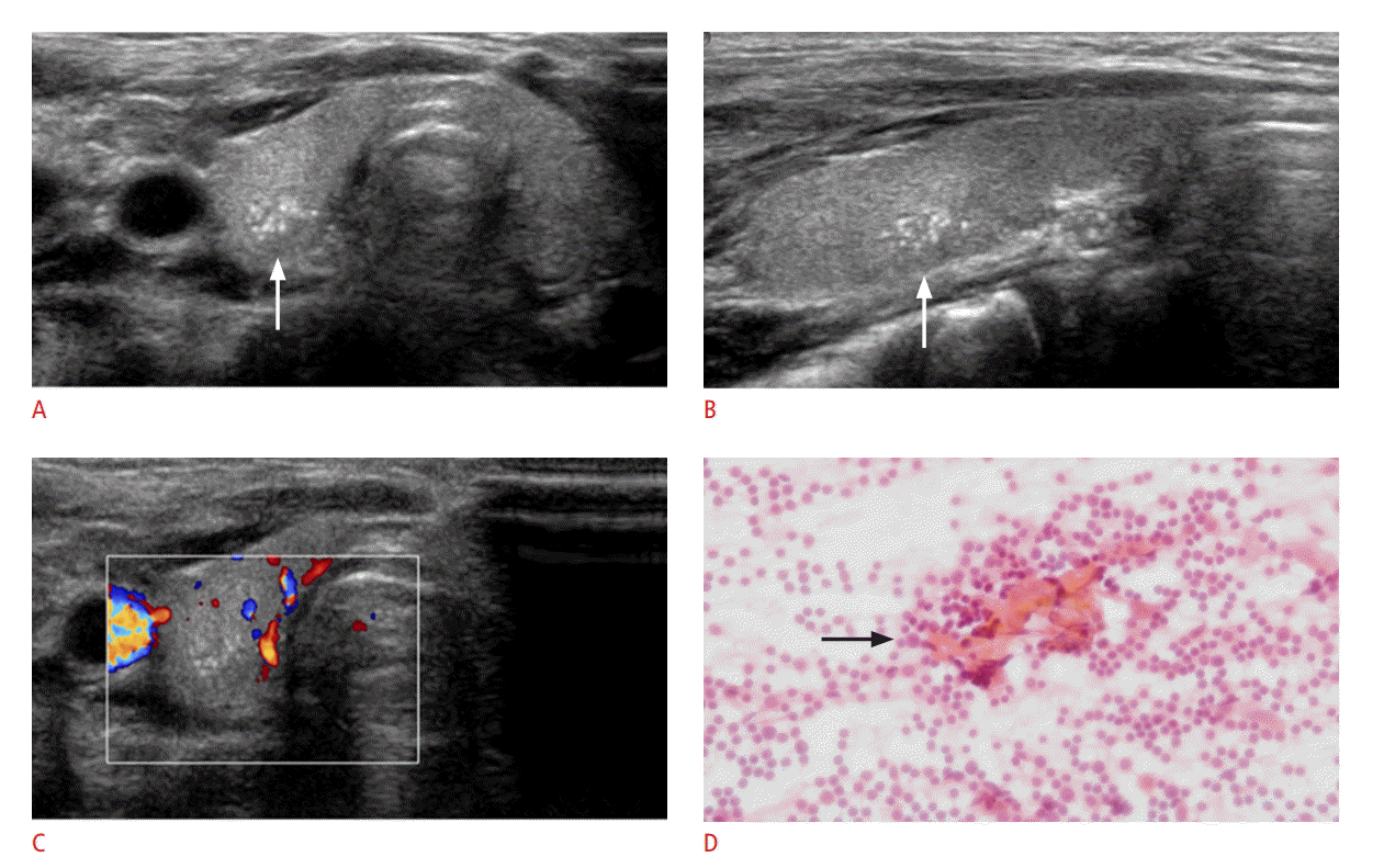
A 4-year-old child with intrathyroidal thymic tissue.
A, B. Transverse (A) and longitudinal (B) scans of gray-scale sonography show an ill-defined nodule (white arrow) with multiple hyperechoic foci in the mid-portion of the right thyroid. C. Color Doppler sonography shows no flow within the nodule. D. A cytological smear slide shows Hassall’s corpuscles (black arrow) with a background of small lymphocytes (H&E, ×400).
Subsequent US-guided fine-needle aspiration biopsy (FNAB) was performed. A cytological investigation revealed Hassall’s corpuscles with a background of small lymphocytes (Fig. 1D) and confirmed the diagnosis of ectopic thymic tissue within the thyroid gland. After 3 months of follow-up by sonography, there were no changes in the size or appearance of the nodule. The sonographic findings of the nodule were similar to those of the thymus.
Discussion
The thymus is embryologically derived from ventral sacculation of the third pharyngeal pouch and minor portions of the fourth pharyngeal pouch in the sixth week of gestation [4]. The bilateral primordial of the thymus starts midline fusion in the eighth week to form the bilobed thymus, which descends into the superior mediastinum [4]. The descent of the thymus and thyroid are closely related because of the proximity of the thyroid diverticulum to the 3rd branchial pouch. During the migration, thymic remnants can be left along the path, which can result in ectopic and accessory thymic tissue in various locations [5]. Examples of this include cervical extension of the thymus, cervical thymus, thymic cyst, cervical thymoma, and ectopic thymus [6]. Ectopic thymic tissue may manifest as a neck mass or an incidentally detected mass.
Thyroid nodules in children are uncommon. The incidence of thyroid nodules in children is about 0.2%-1.5%, compared to about 40%-50% in adults [1,7]. However, thyroid nodules are more often malignant in children, at a rate of 15%-26%, compared to 5%-10% in adults [1,7]. The differential diagnosis for thyroid nodules in children typically includes nodular goiter, lymphocytic thyroiditis, colloid cysts, follicular adenomas, degenerating nodules, and malignant thyroid nodules [3]. In our case, the thyroid nodule showed an ill-defined margin with multiple hyperechoic foci in the mid-portion of the right thyroid. The sonographic findings of multiple hyperechoic foci within the nodule were similar to those seen in thymic tissue. Many reported cases of intrathyroidal thymic tissue have presented as well-defined hypoechoic nodules with linear and dot-like echoes [2]. Intrathyroidal thymic tissue may manifest contiguous with the mediastinal thymus [8]. These sonographic findings are typical of intrathyroidal thymic tissue and can be used to confirm the diagnosis [9]. In our case, however, the nodule had an ill-defined margin. Several sonographic findings that suggest a malignant thyroid nodule are marked hypoechogenicity, irregular margins, microcalcifications, and a taller-than-wide shape [10,11]. We performed a US-guided FNAB to confirm the differential diagnosis of intrathyroidal thymic tissue and thyroid malignancy.
In conclusion, intrathyroidal thymic tissue is rare and may be confused with a malignant thyroid nodule because of hyperechoic dots mimicking calcifications. In children, intrathyroidal thymic tissue should be considered in the differential diagnosis for thyroid nodules. The sonographic findings of ectopic thymus were similar to those of the thymus; therefore, we should be familiar with US findings of normal thymic tissue. If sonography results are inconclusive and further evaluation is needed, a biopsy may be useful for confirmative diagnosis.
Notes
No potential conflict of interest relevant to this article was repoted.