

 Print
Print facebook
 twitter
 Linkedin
 google+

Introduction
Thyroid nodules are very commonly observed on thyroid ultrasonography (US), and conventional US has been widely used to determine which nodules should be biopsied. There are several suspicious US features that predict thyroid cancer, such as hypoechogenicity, marked hypoechogenicity, a microlobulated or spiculated margin, micro- or macro-calcifications, and a taller-than-wide shape [1,2]. Although conventional US can provide meaningful information in thyroid nodule diagnosis, there has been considerable variation in diagnostic performances [2-4].
On physical examination, a hard or firm nature is associated with thyroid malignancy. However, palpation is very subjective and limited in patients with multinodular goiter or small deep-seated nodules [5]. Meanwhile, US-based elastography can provide an objective evaluation of tissue stiffness [6,7]. There are two kinds of elastography (strain and shear wave elastography) that are currently used in clinical practice [8,9]. Although many reports have compared conventional US with elastography, in clinical practice, the final decision or diagnosis is usually based on a combination of conventional US and elastography. This review summarizes the current techniques, diagnostic performance, reproducibility, and limitations of thyroid elastography.
Elastography Techniques
Strain Elastography
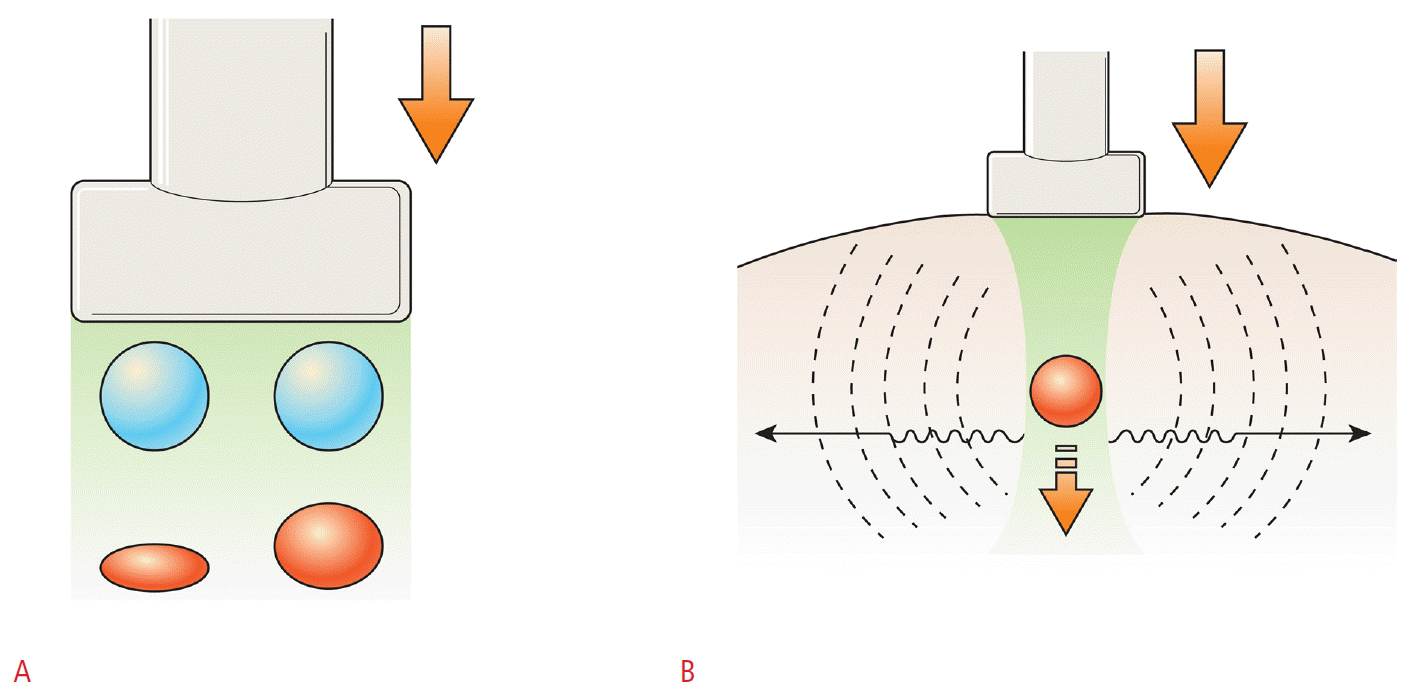
To evaluate the relationship between compression and strain, Young’s modulus, the ratio of pressure to strain, has been used. Strain (or static) elastography requires an external palpation with a probe or endogenous stress such as cardiovascular movements, resulting in an axial displacement of the tissue by mechanical stress (Fig. 1) [10]. Tissue deformation from the stress is measured and visualized in a split-screen mode with a conventional B-mode image and an elastogram on a screen. To acquire the elastographic images, compression is continuously applied by a transducer and followed by decompression. The elastic image is superimposed on the B-mode image, and tissue stiffness is displayed in a continuum of colors from red (soft tissue) to blue (hard tissue). According to the chosen machine, color scales are applied inversely.
By strain elastography, two kinds of elasticity assessments can be obtained. First, visual scoring of colors within and around the nodules can be assessed, using 4-5-scale scoring systems (Fig. 2) [6,7]. Second, two regions of interest (ROIs) are drawn over the target region and the adjacent reference region, respectively. Then, a strain ratio is automatically calculated through the machine. The likelihood of malignancy increases with an increase in the strain ratio [11].
Shear Wave Elastography
Shear waves are the transverse components of particle displacement that are rapidly attenuated by the tissue. Their speed is closely related to Young’s modulus formula, in which tissue elasticity can be assessed from the shear wave propagation speed (Fig. 1) [12]. Shear wave elastography (SWE) provides quantitative elastic information on the basis of the acoustic pulse of a US probe that stimulates tissues, thereby producing a real-time elastogram [13,14]. As SWE is dependent on the production of radiation force by the probe, it is more operator-independent, reproducible, and quantitative. There are two applicable methods for the clinical evaluation of thyroid nodules: the supersonic shear wave and the acoustic radiation force impulse (ARFI) methods [15]. The former uses focused ultrasonic beams that propagate through the entire imaging area. A color-coded image displaying the shear wave velocity (m/sec) or elasticity (kilopascals, kPa) for each pixel in the ROI is acquired. Within a given ROI, a variety of stiffness parameters can be measured, including the mean stiffness (Emean), maximum stiffness (Emax), and standard deviation (SD). Unlike strain elastography, soft tissue is displayed in shades of blue and hard tissue is displayed in red [13,16]. ARFI uses short-duration acoustic pulses that excite the tissue within the ROI. The elasticity is expressed as in meters per second (m/sec) and does not display color-coded images for elastography [17].
Thyroid Malignancy Diagnostic Performance
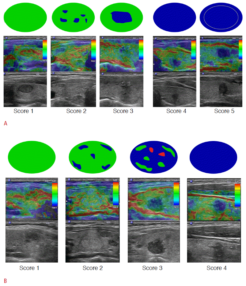
Clinical application of thyroid elastography was first reported in 2007 by Rago et al. [6]; it used five-point scales based on Ueno and Itoh [18]’s study using strain elastography. A score of 1 defined elasticity that is entirely soft in the nodule, 2 as mostly soft in the nodule, 3 as peripherally soft, 4 as entirely hard in the nodule, and 5 as hard in the area under consideration as well as the entire nodule (Fig. 2). Other criteria demonstrated in 2008 by Asteria et al. [7] used four-point scales based on the study of Itoh et al. [10]. Asteria’s criteria defined a score of 1 as elasticity that is entirely soft in the nodule, 2 as mostly soft in the nodule, 3 as mostly hard in the nodule, and 4 as entirely hard in the nodule (Fig. 2) [7]. In their study, nodules with Rago scores of 4 and 5 or Asteria scores of 3 and 4 were regarded as suspicious elastographic features for malignancy. Using Rago’s criteria in a study including 92 consecutive patients with a single nodule, the researchers calculated the sensitivity to be 97% and the specificity to be 100% for predicting thyroid malignancy [6]. Using Asteria’s criteria, the researchers calculated sensitivity and specificity to be 94.1% and 81%, respectively, in 86 nodules [7]. The two investigations evaluated the diagnostic performances of each US feature on gray-scale US but did not evaluate combinations of US features. Further, they did not demonstrate the diagnostic role of a combination of conventional US and elastography. In practice, elastography is usually performed as an extension of conventional US and not as an independent test. Therefore, a comparison of conventional US with elastography can be meaningless in view of its current clinical utility. The value of elastography should be evaluated by comparing conventional US with a combination of conventional US and elastography. In 2012, Moon et al. [19] evaluated the practical role of elastography as an adjunctive tool of gray-scale US in 676 patients with 703 solid nodules. In the study, neither elastography nor the combination of elastography and grayscale US showed better performance for diagnosing thyroid cancers compared with gray-scale US.
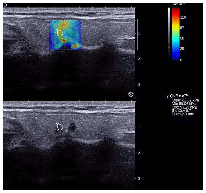
SWE was first reported for diagnosing thyroid nodules in 2010 by Sebag et al. [13] in a study that included 146 nodules form 93 patients. Using significant US features (hypoechogenicity, microcalcifications, and intranodular vascularity) in the study, they calculated the US score by using the following equation: 42.18 (if intranodular vascularity=yes) + 6 (if hypoechogenicity=no) + 30.70 (if hypoechogenicity=yes) + 27.13 (if micro-calcification=yes). The US scores ranged from 0 to 100, with the higher values predicting malignancy. In that study, the best cut-off point for discriminating benign nodules from malignant ones was a mean elasticity index of 65 kPa on SWE and 63.60 (US score) on gray-scale US. The sensitivity and the specificity for malignancy were 51.9% and 97% in the case of gray-scale US and 81.5% and 97.0% in the case of the combination of gray-scale US and SWE, respectively. With these results, they concluded that SWE could be a promising tool for the differentiation of thyroid nodules. However, the sample size was small; further, the calculation of US scores is not commonly used in the clinical field due to its complexity. Thus far, there have been several reports regarding the use of SWE in the differentiation of benign and malignant nodules. Bhatia et al. [20] investigated the role of SWE using several parameters such as the mean elasticity index of the largest ROI within the whole lesion, the mean elasticity index of a 2-mm-diameter area in the stiffest region, the maximum and minimum elasticity index of the largest ROIs within the whole lesion, and standard deviation of the largest ROIs within the whole lesion (Fig. 3). Among these numerous parameters, the most accurate value was that of the mean elasticity index of a 2-mm-diameter area in the stiffest region with a cut-off value of 34.5 kPa. The sensitivity and the specificity for predicting malignancy were 76.9% and 71.1%, respectively. Veyrieres et al. [21] also evaluated the diagnostic performances of SWE in the case of 297 thyroid nodules. The sensitivity and the specificity had good results of 80% and 90.5%, respectively, with the best cutoff value of 66 kPa. They measured the elasticity index of ROIs covering the visually stiffer nodule regions on elastographic color mapping, which resulted in differently sized ROIs according to the nodules. Kim et al. [14] studied the role of SWE when combined with conventional US. They used several parameters of SWE, including the mean elasticity index of the stiffest portion of mass or surrounding tissue, the maximum elasticity index of the stiffest portion of mass or surrounding tissue, the minimum elasticity index of the stiffest portion of mass or surrounding tissue, the ratio of the mean elasticity score of the lesion and parenchyma, and the ratio of the mean elasticity score of the lesion and the strap muscle. Among them, the maximum elasticity index of the stiffest portion of mass or surrounding tissue had the highest areas under the ROC curves. Although the researchers concluded that the combination of quantitative SWE and conventional US had higher specificity than conventional US alone for predicting malignancy, the areas under the ROC curves were higher in conventional US alone than in the combination of quantitative SWE and conventional US [14]. The most recent report of SWE was presented by Szczepanek-Parulska et al. [22]. They evaluated the diagnostic powers of each US feature of conventional US and SWE both qualitatively and quantitatively in the case of 393 thyroid nodules. However, in their study, they did not definitely document their approach to finding the elasticity index. The best cutoff elasticity index providing the highest odds ratio was 50 kPa, and a qualitative evaluation of SWE was not inferior to a quantitative evaluation of thyroid lesions.
Thus far, the abovementioned results are considered controversial despite many reports on the diagnostic utility of thyroid elastography (Table 1) [14,19,21,23-28]. Several investigators have compared elastography with each suspicious US feature and not with a combination of suspicious US features on conventional US [6,29]. Considering that the final assessment of a thyroid nodule is usually made on the basis of variable US features, the comparison of each suspicious US feature to elastography can only provide limited information for physicians [1,2]. For elastography to be an excellent adjunctive tool to conventional US, the combination of conventional US and elastography should have higher accuracy and sensitivity than conventional US alone. Most reports have stated that a combination of conventional US and elastography showed higher sensitivity than conventional US alone. In contrast, the diagnostic accuracy, specificity, and positive predictive value were inferior to those of conventional US alone. Therefore, the clinical application of elastography should be decided by how much experience the operator has with both conventional US and elastography for the thyroid.
Diagnostic Utility in the Presurgical Diagnosis of Thyroid Nodules with Nondiagnostic or Indeterminate Cytology
US-guided fine-needle aspiration (US-FNA) has been widely used to diagnose thyroid nodules with excellent performance and less invasiveness. It has been considered the standard diagnostic tool to diagnose a thyroid nodule [30,31]. However, US-FNA has major limitations, such as “indeterminate” or “non-diagnostic” cytology, which can occur for up to 30% of all aspirated thyroid nodules [32,33]. To overcome these limitations, there have been several methods such as molecular markers and intraoperative frozen section [34-36]. The former need additional costs and the latter can be performed intraoperatively, not preoperatively.
Some investigators have attempted to evaluate the role of thyroid elastography in thyroid nodules with “indeterminate” or “nondiagnostic” cytology. The first study was performed in 32 patients with thyroid nodules with “indeterminate” cytology by Rago et al. [6]; it showed better diagnostic performance for each suspicious US feature. The two subsequent studies demonstrated the usefulness of thyroid elastography to predict thyroid cancer using qualitative and quantitative methods [37,38]. However, Lippolis et al. [39]’s study showed the opposite result; the study considered 102 thyroid nodules with “indeterminate” cytology and used qualitative elastography. The above studies evaluated the diagnostic utility of conventional US in a thyroid nodule with “indeterminate” cytology using each US feature, not a combination of US features. Although there were two studies concerning the value of thyroid elastography in thyroid nodules with “non-diagnostic” cytology [27,38], conventional US also showed good diagnostic results in thyroid nodules with “non-diagnostic” cytology even without additional time and cost [33]. Therefore, further validation is needed in the field.
Reproducibility
To overcome the subjectivity of palpation on physical examination, elastography has been developed to objectively evaluate tissue firmness or hardness. However, elastography can be affected by an operator’s degree of compression and experience. Park et al. [5] were the first to report on the interobserver agreement of strain elastography of thyroid cancers. They independently examined thyroid cancers by three operators, using conventional US and strain elastography. The agreement was very poor in the case of strain elastography, in contrast to a better interobserver agreement in the case of conventional US. Park et al. [5] speculated that different external compressions by the probe and carotid artery pulsation influenced the poor agreement. In their study, the elastographic machine did not have any objective parameters to show the compressive force generated by the probe. Since then, there have been several reports on the reproducibility of elastography (Table 2) [5,11,15,20,21,40-43]. Most of them show substantial or almost perfect agreement. The remarkable improvement in interobserver agreement from the study by Park et al. [5] can be explained as follows: First, strain elastographic machines have the ability to monitor compressive force using real-time monitors, thereby reducing the over- or under-compression by different operators that previously influenced elastographic scoring [40]. Second, SWE has different physics from strain elastography, resulting in reproducible results [13,14].
Limitations
Several factors can affect the results of elastography, including nodule characteristics (calcifications and cystic components), the experience of the operator, and motion artifacts such as carotid artery pulsation [40,44]. To overcome these limitations, elastography can be selectively used in thyroid nodules without calcifications and cystic changes, and should be performed by experienced operators using objective parameters provided by elastographic machines.
With respect to SWE, there have been no definite cut-off values given for the elasticity index even when the same machine was used, and no method has yet been standardized for the measurement of the thyroid lesion areas [13,14,20-22]. Further studies are needed to provide a practical guideline for the evaluation of thyroid nodules with SWE.
Conclusion
While elastography is a promising technique in some organs such as the breast [45,46] and the liver [47-50], there have been conflicting results of its additional value in predicting thyroid malignancy [14,19-26]. To be a good adjunctive diagnostic tool to conventional US, additional elastography should improve the diagnostic performances of conventional US, rationalizing the consequently longer US time and the additional cost of elastographic software that comes with its use. The value of elastography may be limited in institutions that show high diagnostic performances of conventional US by highly dedicated physicians. However, further studies should continuously validate the utility of elastography, particularly in selected cases.
 Download Citation
 PDF Links
 PubReader
 ePub Link
 Full text via DOI
 Full text via PMC


