Introduction
With improvements in the technology of ultrasonography, color Doppler ultrasonography (CDUS) has come to exhibit great utility for imaging superficial structures, since submillimeter-scale anatomic structures and blood flow in tiny vessels can be investigated in real time. The penis is a protruding superficial organ that is composed of many different types of soft-tissue structures. Additionally, it is a highly vascularized organ, making CDUS imaging suitable for the penis. CDUS enables the excellent evaluation of normal and pathologic penile conditions with optimal accuracy in real time. In the 1990s, CDUS became an essential tool for the identification and classification of organic causes of erectile dysfunction (ED). However, the introduction of an effective oral medication (phosphodiesterase type 5 inhibitor; PDE5-I) has markedly reduced the utility of CDUS in ED patients. Poor long-term clinical outcomes of surgery for ED were another reason that the use of CDUS declined. Even though the importance of CDUS has decreased, it has maintained a certain role for the evaluation of non-responders to PDE5-I [1,2].
Recently, as the survival rate of cancer patients after pelvic surgery for prostate, bladder, or rectal cancer has improved, postoperative ED is becoming an important issue in cancer survivors with respect to quality of life. In these cases, penile CDUS is an efficient and convenient tool for the assessment of postoperative ED [3]. Additionally, penile CDUS has come to play an increasing role in the detection of silent coronary artery disease (CAD) in men presenting with ED. ED is now recognized as one of the earliest manifestations of endothelial dysfunction and peripheral vascular disease [4]. A Doppler workup of the penile vessels can be used to determine the need for further cardiac assessments in high-risk patients with CAD, and it can be used as a noninvasive method for evaluating endothelial dysfunction. Recent developments in elastography and contrast-enhanced ultrasonography techniques might help provide valuable information on penile pathology [5,6].
Ultrasonographic Anatomy of the Penis
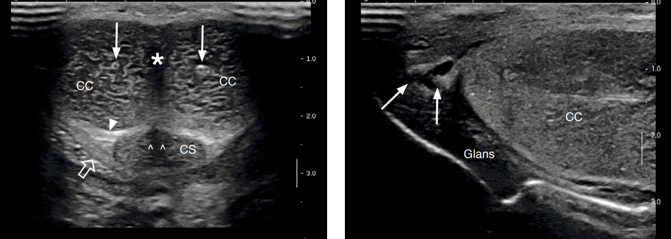
The penile body consists of two paired corpora cavernosa, which are the main erectile bodies, and the corpus spongiosum, which contains the urethra (Fig. 1). The outer dartos fascia and the inner Buck fascia contain the deep dorsal vein (DDV) and a paired dorsal neurovascular bundle. The tunica albuginea (TA) also has two layers: an outer layer of longitudinal fibers and an inner layer of circular fibers with perpendicular struts/pillars, which serve to augment the support provided by the intracavernosal septum. The inner portion of the corpora consists of interconnected sinusoids separated by smooth muscle trabeculae. The corpus spongiosum lacks the outer longitudinal layer, as well as strut structures [7].
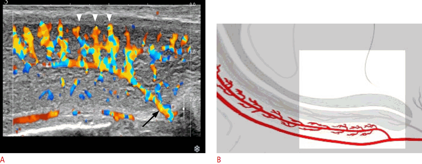
The primary source of blood supply to the penis is usually through the internal pudendal artery, which arises from the anterior division of the internal iliac artery. The internal pudendal artery becomes the common penile artery after giving off a branch to the perineum. The branches of the penile artery are the dorsal artery, the bulbourethral artery, and the cavernosal artery (CA). The CA is responsible for the tumescence of the corpus cavernosum, and the dorsal artery is responsible for the engorgement of the glans penis during erection. Distally, three branches of penile arteries join to form a vascular ring, as an anastomosis, near the glans. The CA gives off many helicine arteries, which supply the trabecular erectile tissue and the sinusoids (Fig. 2). The helicine arteries are contracted and tortuous in the flaccid state and become dilated and straight during erection. Venous drainage from the three corpora originates in the tiny venules just beneath the TA, forming the subtunical venular plexus before exiting as the emissary veins. Outside the TA, a prominent DDV is the main venous drainage via the circumflex vein. Then, a single DDV runs upward behind the symphysis pubis to join the periprostatic venous plexus [8].
Penile Erection on CDUS
Penile erection is primarily a neurovascular event. Sexual stimulation causes a release of neurotransmitters from the cavernous nerve terminals and of relaxing factors from the endothelial cells, resulting in relaxation of the smooth muscle in the arterioles. A severalfold increase in blood flow with concomitant relaxation of the cavernous smooth muscle facilitates the rapid filling and expansion of sinusoids against the TA. Therefore, the sub-tunical venular plexuses are compressed, resulting in the almost complete occlusion of venous outflow. After that, blood is trapped within the cavernosa, which raises the intracavernosal pressure (ICP), corresponding to full erection. During sexual activity, the ischiocavernosus muscle compresses the blood-filled base of the corpus cavernosum. The penis enters the rigid erection phase, during which inflow and outflow temporarily cease. Detumescence starts with contraction of the trabecular smooth muscle and opening of the venous channel, expelling the trapped blood and restoring flaccidity [9].
Penile ultrasonography (US) should be performed using high frequency linear probes with the patient in the supine position. The penis is scanned from its ventral surface using longitudinal and transverse views. Evaluation should be carried out while the penis is flaccid and after the intravenous injection of vasoactive drugs. CDUS with spectrum analysis is performed with tuning for a slow-flow setting. A mixture of agents such as papaverine, phentolamine, and prostaglandin E1 is injected into the lateral aspect of the midshaft of the penis, after evaluation of the flaccid state. Penile US requires some basic rules of privacy and a private environment. In addition, the possibility of complications, including priapism (an erection that lasts for more than 4 hours), hypotension, pain, and hematoma, after vasoactive stimulation should be explained to the patient before injection [10].
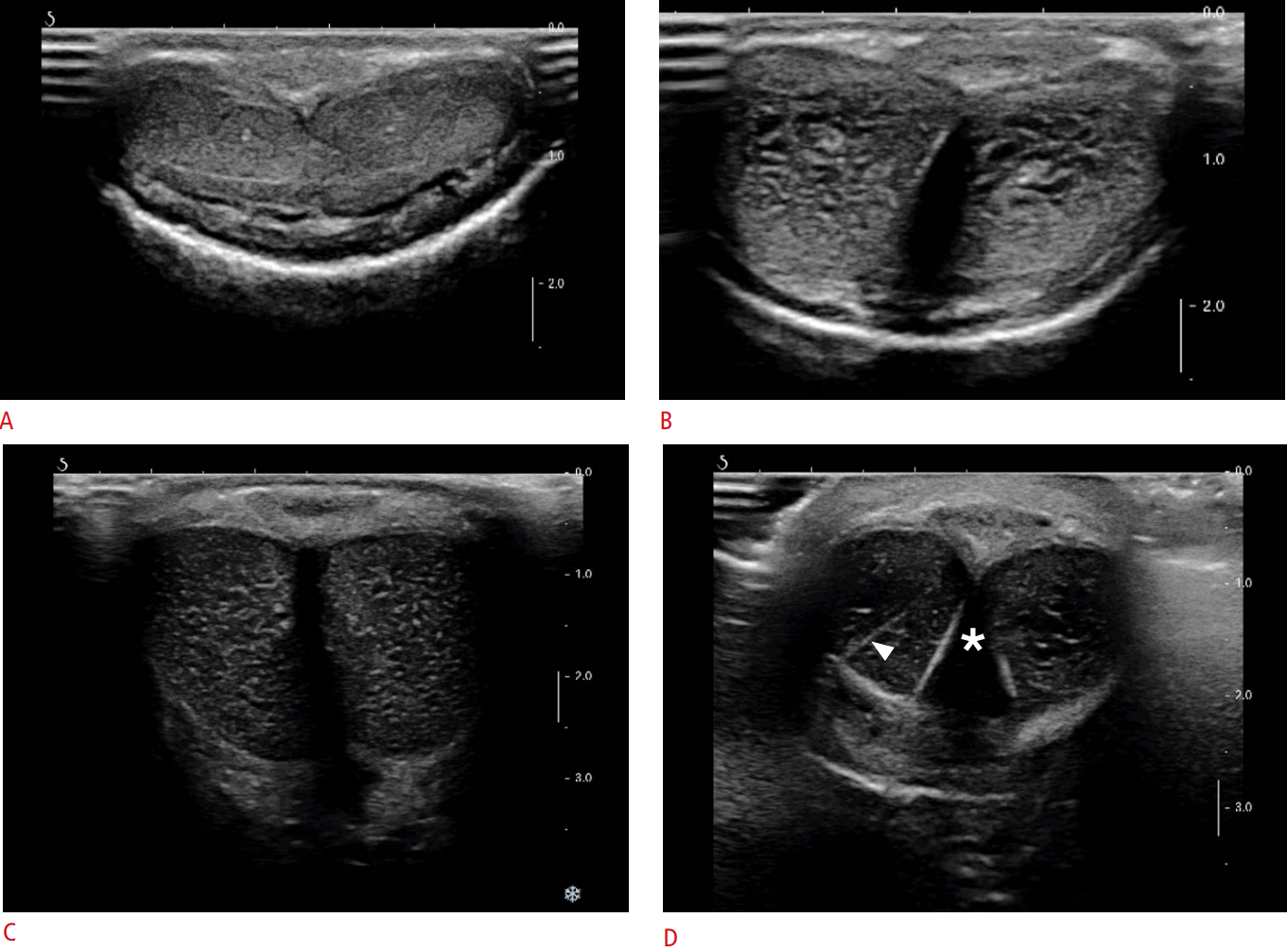
In the flaccid state, penile bodies present with intermediate echogenicity and homogeneous echotexture. After a cavernosal injection, sinusoidal distension begins in the central portion of the cavernosa, which appears less echogenic than the outer portion. During the filling phase, we can see the fine echogenic network in the corpus cavernosum due to numerous sinusoidal interfaces (Fig. 3). The penile septum appears as an echogenic structure with back attenuation, which can hamper the evaluation of the dorsal aspect of the penis and the TA [9].
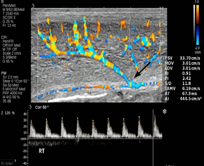
The peak systolic velocity (PSV) of the CA varies significantly according to the sampling location and angle. In order to standardize the velocity measurements, spectral sampling of the CA is obtained at the origin, on the base of the penis under the guidance of the color Doppler signal, where the CA angles posteriorly toward the crus. It is preferable to apply the angle correction cursor or the steering box (Fig. 4). Fig. 5 shows normal waveform changes in the CA during erection. In the flaccid phase, monophasic waveforms are recognized in the CA with a low-velocity, high-resistance flow (only systole without diastole). After the onset of erection, an increased systole and diastole, corresponding to the beginning of the filling phase, is noted. When the ICP begins to rise, a dicrotic notch appears at the end of systole, and progressively decreased diastole is observed. As the ICP equals the systemic diastolic pressure, the end diastolic velocity (EDV) disappears. Then, diastole is reversed due to the increased ICP above it, reflecting the full erection phase. During rigid erection, reduction in the PSV and disappearance of diastole are seen. The rigid erection phase is not commonly visible after an induced erection. A time-velocity curve can be used to illustrate hemodynamic changes in the different erectile phases. The period between the starting point of decreasing diastolic flow and reversed diastole indicates the period in which the veno-occlusive mechanism is operative. Since dorsal arteries are outside the TA, and therefore they are not subject to ICP changes and the erection phase, they show persistent antegrade diastolic flow. Dorsally, the midline-located DDV is not consistently visible in normal subjects during all erection phases as well as in the flaccid phase. However, it can be identified on detumescence. A prominent DDV is commonly visualized in veno-occlusive ED [11].
Erectile Dysfunction
ED is classified as psychogenic, organic (neurogenic, hormonal, vasculogenic, and drug-induced), and mixed. Mixed ED is the most common, and involves both a psychogenic and an organic component. Using CDUS, we can identify and classify the organic component of ED. Based on the findings on Doppler US, ED can be categorized into arterial and venous types. Arteriogenic ED involves impairment of the arterial influx into the cavernosum due to various causes. In contrast, an impaired veno-occlusive mechanism causes venous leak, resulting in what is known as venogenic ED. PSV is the most accurate indicator of arterial disease. Arterial insufficiency is diagnosed when the PSV is less than 25 cm/sec, with 92% accuracy. Veno-occlusive ED shows a persistent EDV over 5 cm/sec during all phases of erection, and flow in the DDV is visible on Doppler US during all phases (Fig. 6). The diagnosis of mixed ED cannot be made using Doppler ultrasonography because venous competence cannot be assessed in a patient with arterial insufficiency. In practice, most elderly patients or postoperative ED patients present with this indeterminate form [12-14].
Although the anatomical and structural changes on gray-scale US of the cavernosa are informative, the functional characteristics after injection are more important. Diagnosis is usually made during scanning in real time based on structural and functional data. Before performing Doppler US, we should review the patient’s self-reported replies to questionnaires and event logs of erectile function, such as the International Index of Erectile Function. In practice, a simplified and reduced version is used [15].
PSV and EDV are the major Doppler parameters for diagnosing ED. The change in the diameter of the CA and the flow signals of DDV as well as the resistive index (RI) are minor parameters for differentiating between the types of ED. Table 1 summarizes the various criteria for the major and minor parameters. A PSV of 25 cm/sec is a key indicator of arterial insufficiency. Additional, diastolic flow reversal can indicate an intact veno-occlusive mechanism. Among the minor criteria, DDV is used as an adjuvant for venous leak. The cavernosal diameter and RI values are also useful, but they have a wide variation and are not repeatable.
Priapism and Peyronie Disease
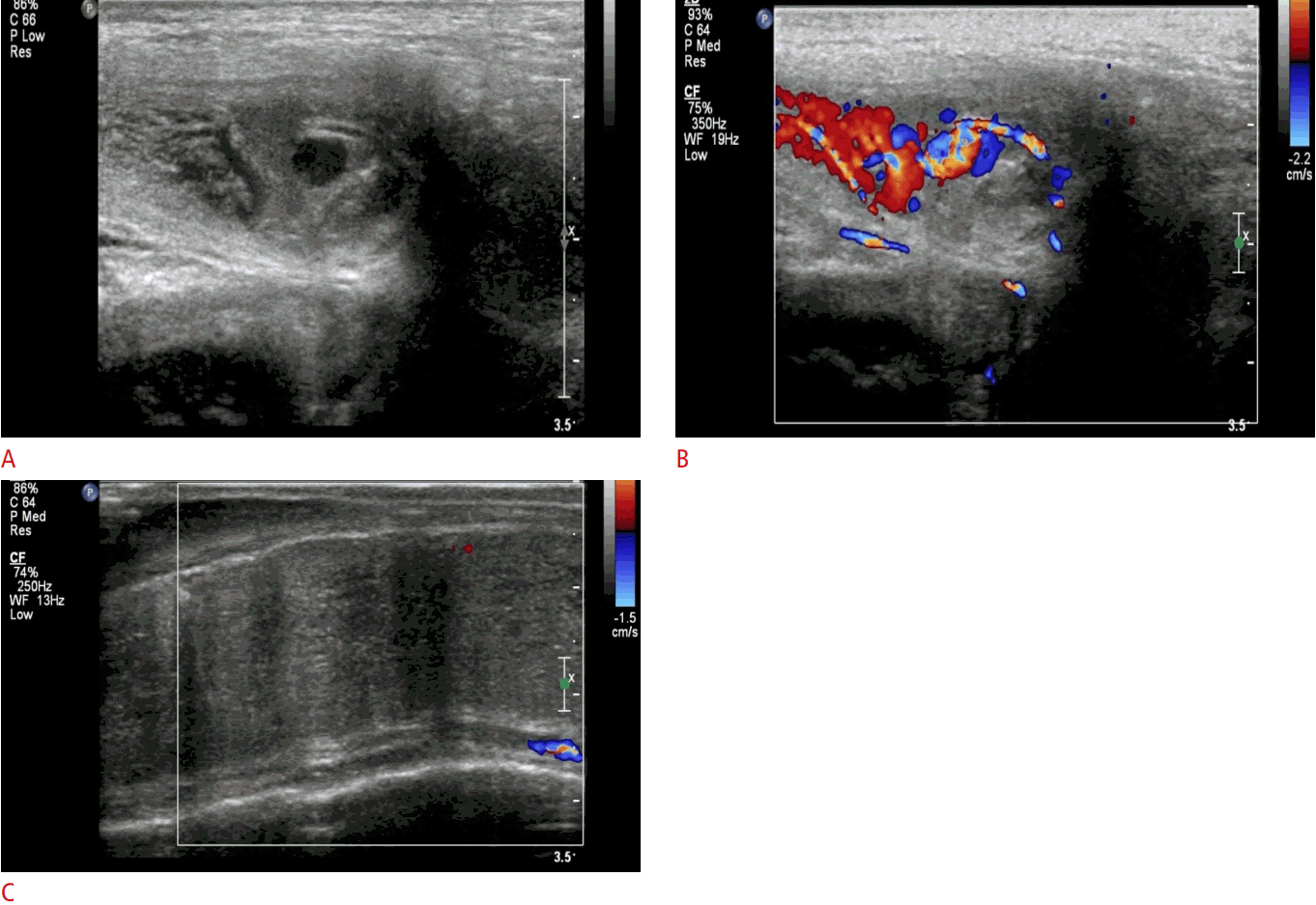
Priapism is defined as persistent tumescence unrelated to sexual stimulation, and it is classified into arterial and venous types. Venous or ischemic priapism is more common than the arterial type, and it is a medical emergency that is caused by decreased or absent venous drainage. Arterial or high-flow priapism is secondary to an uncontrolled increased arterial influx in cases of fistula formation or cavernosal metastasis of a solid tumor. The arterial-lacunar fistula in high-flow priapism bypasses the helicine arteries, directly extending into the cavernosal tissue and appearing as a characteristic color blush and turbulent high-velocity flow on Doppler analysis (Fig. 7). Low-flow or ischemic priapism is characterized by the absence of cavernosal blood flow and the failure of detumescence. On CDUS, low-flow priapism usually presents with a lack of CA blood flow or with a very high-resistance flow pattern in the CA (Fig. 7) [16,17].
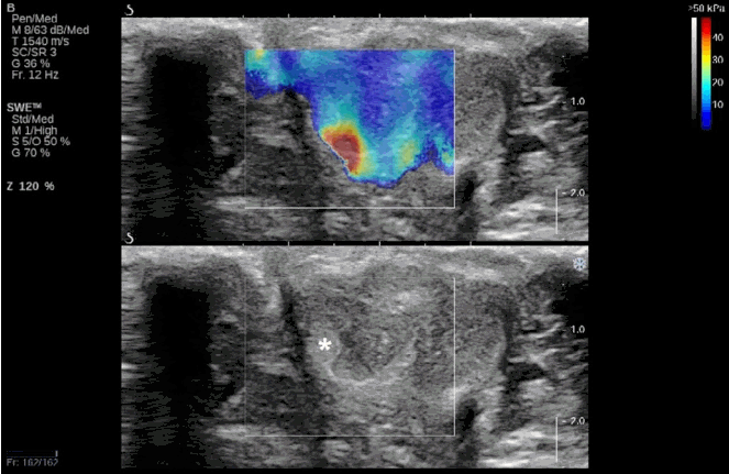
Peyronie disease is characterized by progressive curvature and shortening of the penile shaft, and may be associated with a palpable nodule, eventually leading to the development of pain during erection and dyspareunia. Gray-scale penile US shows multiple calcified nodules along the tunical envelope of the cavernosum, associated with veno-occlusive ED. Fibrous thickening or a plaque without calcification is missed on gray-scale US. The recently developed technique of sonoelastography is helpful for differentiating and detecting noncalcified fibrous plaques (Fig. 8) [5,18,19].
Current Status of Penile CDUS
The importance of penile Doppler US has diminished over the last 2-3 decades because of the poor long-term clinical outcomes of surgery as well as the introduction of oral PDE5-I. PDE5-I revolutionized ED management, as a high-quality erection after PDE5-I confirms adequate arterial inflow and effective veno-occlusive mechanisms. However, penile CDUS has continued to play a role in the evaluation of non-responders to PDE5-I and in the assessment of cancer patients with ED after pelvic surgery due to prostate or rectal cancer. Additionally, penile CDUS has come to play an increasing role in the detection of silent CAD in men presenting with ED. ED is now recognized as one of the earliest manifestations of endothelial dysfunction and peripheral vascular disease. A Doppler workup of the penile vessels in patients who have significant risk factors for cardiovascular disease can be used to assess the need for further cardiac or peripheral vascular assessments. Continuous improvements in real-time spatial resolution and contrast have broadened the indications for penile CDUS, such as preoperative evaluation for Peyronie disease and the detection and quantification of penile fibrosis. Additionally, the recent advent of sonoelastography and contrast-enhanced ultrasonography is also expected to provide valuable information on penile pathology, and much research is being conducted into these possibilities [3,20,21].
Unfortunately, only a limited number of specialists can currently perform penile CDUS, while many clinicians, including radiologists, often have a superficial, insufficient, or obsolete knowledge of this modality. Becoming familiar with the latest knowledge in the field of penile CDUS is essential for many patients with variable penile pathologies to be successfully and conveniently assessed with CDUS. Familiarity with these quantitative parameters and the imaging features of penile CDUS may aid in the detection and classification of penile pathologies, resulting in the facilitation of appropriate management.


 Print
Print facebook
 twitter
 Linkedin
 google+


 Download Citation
 PDF Links
 PubReader
 ePub Link
 Full text via DOI
 Full text via PMC


