

 Print
Print facebook
 twitter
 Linkedin
 google+

Introduction
Gross hematuria in children is related to a wide spectrum of conditions, and the majority of cases have a relatively benign etiology compared to cases in adults [1]. Major causes of gross hematuria in children include infection, trauma, metabolic diseases, autoimmune diseases, and glomerulonephropathies [1]. Pediatric neoplasms in the urinary system are rare due to the relatively low incidence of epithelial tumors. Childhood bladder hemangiomas, among them, are seldom reported due to their extreme rarity [2]. In the present case, we describe multiple urinary bladder hemangiomas as a cause of pediatric gross hematuria focused on ultrasonographic findings.
Case Report
The Institutional Review Board of our hospital approved this case study and waived the requirement for informed consent. A 4-year-old boy visited our outpatient clinic with recurrent painless gross hematuria with blood clots. His urine color deepened at the end of micturition, reflecting disease of the bladder. Hematuria continued for several days and had an intermittent and recurrent pattern of presentation. The child had no significant medical or family history. Vital signs and observations from the physical examination were within normal limits. The blood cell count results were as follows: hemoglobin level, 12.6 g/dL; hematocrit, 39.4%; and platelet count, 347,000/╬╝L. Random urinalysis with microscopic examination showed hematuria with a negligible count of dysmorphic red blood cells (<5%), without proteinuria or pyuria.
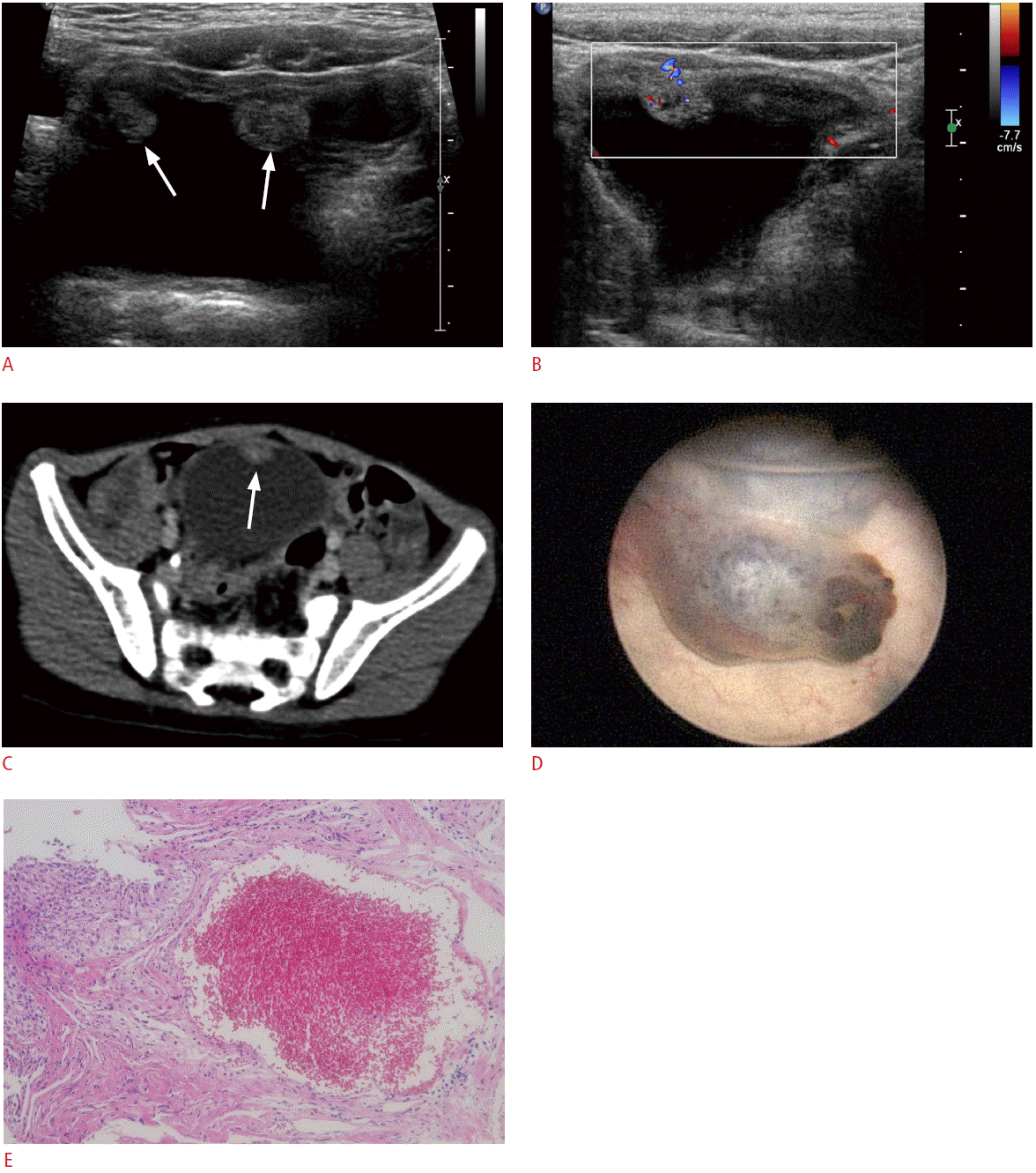
The patient underwent urinary system ultrasonography to evaluate the hematuria. Pelvic ultrasonography images (Fig. 1A) revealed multiple intraluminal polypoid lesions of variable sizes from 0.9 cm to 1.3 cm in the urinary bladder. The lesions were dispersed along the bladder wall and not confined to the bladder dome. There was no definite muscular layer involvement or perivesical infiltration observed on ultrasonography. Vascularity of the polypoid lesions was mildly increased on Doppler ultrasonography (Fig. 1B). There was no bladder wall thickening or prominent trabeculation to suggest cystitis. Both kidneys were grossly normal without evidence of hydronephrosis or urinary stones. Pelvic computed tomography (CT) with contrast enhancement was also performed to evaluate the extent of the lesions and pelvic lymphadenopathy. The CT images showed an enhancing intraluminal polypoid mass on the bladder wall, without visible calcification or perivesical invasion (Fig. 1C). These imaging findings suggested the possibility of a benign bladder tumor without specific differential diagnosis.
For further evaluation of the bladder lesions, a cystoscopic examination was performed. Blue to reddish sessile lesions of various sizes were visualized on the bladder dome and along the lateral aspects of the urinary bladder (Fig. 1D). Blood vessels covered the adjacent bladder mucosa in a reticular pattern. The urethra and bilateral ureteral orifices were not remarkable. Cold-cup biopsy was conducted at the bladder dome lesion, and the remaining portions were coagulated with a Holmium laser. Pathologic examination revealed findings of large cystically dilated vessels with thin walls in the submucosa, consistent with cavernous hemangioma (Fig. 1E). Physical examination revealed no other skin or palpable lesions suggestive of hemangioma. Postoperative pelvic ultrasonography showed that the sizes of the isoechoic intraluminal lesions in the urinary bladder had decreased. Hematuria was not noted on postoperative urinalysis.
Discussion
Gross hematuria in children is a relatively uncommon manifestation, accounting for about 1 in 1,000 outpatient visits. Even though approximately half of cases with gross hematuria remain idiopathic, identification of the underlying cause is important for appropriate management [1]. Among the various possible causes of hematuria in children, urinary tract tumors are rare. Neoplasms of the bladder account for a very small proportion of known etiologies. Greenfield et al. [1] reviewed gross hematuria in 342 children and reported that only 1% of cases were associated with urinary tract neoplasms, including low-grade transitional cell carcinoma and Wilms tumor.
Even though it is rare, hemangioma may be the most common benign bladder tumor found in children [3]. It usually occurs in children and young adolescents and most commonly presents with painless recurrent gross hematuria. However, hypovolemic shock can be present in cases with massive hemorrhage [4]. This tumor may occur sporadically or as a component of a syndrome such as Sturge-Weber or Klippel-Trenaunay-Weber syndrome [5]. Approximately 30% of bladder hemangiomas are associated with additional hemangiomas in other parts of the body [6], although this was not observed in our case. Three histologic subtypes of hemangioma have been characterized, with the cavernous form being the most common, followed by the capillary and arteriovenous subtypes [7]. The histologic depth of a bladder hemangioma may be confined to the submucosa, although extension to the muscular layer or even to the perivesical tissues is common [4]. Tumors are most frequently located in the bladder dome, on the posterolateral walls, or in the trigone [4,5]. The basis of management is ablation of the lesion. For large lesions or those that extend deep into the bladder, open resection of the lesion or partial cystectomy may be necessary [4].
Ultrasonography is the first-line imaging modality for evaluation of pediatric gross hematuria, as it does not involve ionizing radiation or require sedation, while producing images relatively quickly and painlessly. Kogan et al. [5] described two forms of bladder hemangioma according to the extension into the bladder wall, a well-marginated intraluminal solid mass and a diffuse bladder wall thickening with punctate calcifications. The mass can be either hyperechoic or hypoechoic. Our case showed intraluminal masses that were isoechoic compared to the bladder wall, without any thickening of the bladder wall. Bladder hemangiomas may have increased vascularity on Doppler ultrasonography [5]. Hydroureteronephrosis can occur as a result of ureteric obstruction by the mass, and a hematoma can obscure the mass in the bladder when there is massive bleeding [4].
Most cases of bladder hemangioma are solitary. Cheng et al. [7] reviewed bladder hemangiomas reported over 60 years and found only two cases with multiplicity among 19 adult patients. The sizes varied from 0.5 cm to 10 cm, although the median size was 0.7 cm, and most lesions were smaller than 3 cm [7]. Our case was typical in respect to size, with lesions ranging from 0.9 cm to 1.3 cm in diameter, although the characteristic of multiplicity was not usual.
Differential diagnosis of a polypoid bladder mass detected in children with painless gross hematuria includes not only hemangioma but also rhabdomyosarcoma, other vascular tumors, inflammatory pseudotumor, leiomyoma, neurofibromatosis, pheochromocytoma, transitional cell papilloma, transitional cell carcinoma, and pseudotumoral cystitis. Rhabdomyosarcoma is the most common tumor arising from the bladder and can present as a polypoid mass. Various vascular tumors of the bladder can also cause hematuria, although these are rare in children with only a few case reports [8]. Inflammatory pseudotumor of the bladder commonly appears as a polypoid mass or a submucosal nodule, with ulceration and hemorrhage [9]. Leiomyoma arises from the submucosal or muscular layer of the bladder and presents as a well-marginated homogenous solid mass on ultrasonography [10]. Bladder involvement of neurofibromatosis is rare and usually manifests as a generalized neurofibromatosis rather than an isolated form [2]. It usually shows diffuse and irregular wall thickening with or without nodules. Bladder pheochromocytoma typically appears as a well-demarcated soft tissue mass with or without a hemorrhagic component, and can be suspected in a patient with adrenergic symptoms [11]. Transitional cell papilloma can appear as a papillary mass on the urinary bladder wall in a child with painless gross hematuria [12]. Although it is very rare, transitional cell carcinoma can also occur in children, showing an irregular papillary appearance or diffuse bladder wall thickening on ultrasonography [1]. Pseudotumoral cystitis, an uncommon form of cystitis, presents with hypoechoic or hyperechoic mass-like elevations of the mucosal surface, mimicking a bladder neoplasm on ultrasonography. Although these various benign and malignant tumors may appear as intraluminal polypoid bladder masses on imaging studies [2], hemangioma should be considered in children when the mass has a benign-looking appearance such as a well-defined margin and no extravesical extension, while showing vascularity despite its small size and multiplicity.
In conclusion, urinary bladder hemangioma should be included in the differential diagnosis of intravesical polypoid mass on ultrasonography in children with gross hematuria, especially when the mass shows a well-defined margin, vascularity despite its small size, and multiplicity.
 Download Citation
 PDF Links
 PubReader
 ePub Link
 Full text via DOI
 Full text via PMC


