Ultrasonography of acute flank pain: a focus on renal stones and acute pyelonephritis
Article information
Abstract
Ultrasonography is a useful tool for the differential diagnosis of acute flank pain. Renal stones appear as a focal area of echogenicity with acoustic shadowing on ultrasonography. In acute pyelonephritis (APN), the kidneys may be enlarged and have a hypoechoic parenchyma with loss of the normal corticomedullary junction. However, clinical and laboratory correlations are essential for the diagnosis of renal stones and APN through imaging studies. This review describes the typical ultrasonography features of renal stones and APN. Moreover, in daily practice, cross-sectional imaging is essential and widely used to confirm renal stones and APN and to differentiate them from other diseases causing flank pain. Other diseases causing acute flank pain are also described in this review.
Introduction
Acute flank pain due to renal stones or acute pyelonephritis (APN) is a common problem in patients presenting to emergency departments. Radiology plays a vital role in the evaluation of these patients. Several imaging modalities can be used, including ultrasonography (US), computed tomography (CT), conventional radiography, and intravenous urography [1,2]. The classic presentation of renal colic is the acute onset of unilateral flank pain with radiation to the groin, dysuria, and hematuria. US can be used to diagnose stones in the renal collecting system when a focal area of echogenicity with acoustic shadowing is identified. When examining the right kidney, the liver can be used as a sonic window, with the patient tilted 15° to the left. When examining the left kidney, the left lateral decubitus position is helpful. In general, directing the patient to breathe deeply and hold his or her breath for a while will help move the ribs and bowel gases out of the window. As in other US examinations, a high-frequency transducer should be used as long as sufficient penetration is achieved in the US of the kidney, and a convex probe of about 5 MHz is usually used in adults. In order to effectively detect renal stones on US, the ultrasound gain should be adjusted to be slightly lower and the focal zone should be adjusted to be near the suspicious stones. If the focal zone is located slightly deeper than the stones, posterior shadowing is more pronounced and less noise is present within the window.
Associated dilatation of the renal collecting system may occur, depending on the size and location of the stone, the length of time the obstruction has been present, and whether the obstruction is partial or complete. If hydronephrosis is suspected, it is necessary to check the fullness of the bladder. If the bladder is full, the kidney should be rechecked after urination to confirm whether the hydronephrosis is due to an overdistended bladder.
In APN, the ultrasonographic appearance of the kidney is usually normal-as are the CT findings of the kidney-but the kidney may be enlarged and have a hypoechoic or hyperechoic parenchyma with loss of the normal corticomedullary junction, abscess formation, or areas of hyperperfusion on color Doppler images [3].
Although US is useful for the diagnosis of renal stones and APN, CT is the main imaging tool that is used, as it provides highly specific findings and an accurate assessment of the renal and extrarenal extent of disease. The purpose of this review is to discuss practical approaches, the findings of US evaluations of acute flank pain, and the pitfalls of US.
Clinical Considerations Regarding Renal Stones
Renal or ureteric colic is the most common urologic emergency and one of the most common cause of acute abdominal pain. Patients treated for renal stones are usually between 30 and 60 years of age. The disease affects men 3 times as often as it does women [2]. Renal colic manifests as the sudden onset of severe pain radiating to the flank or inguinal area. This severe pain is caused by the passage of a stone formed in the kidney [4]. The passage of stones into the ureter with subsequent acute obstruction, proximal urinary tract dilation, and spasm is associated with classic renal colic. The most useful laboratory finding for renal stones is hematuria on microscopic urinalysis. However, the specificity (48%) and negative predictive value (65%) of this finding were found to be low [5]. Thus, imaging studies are essential for diagnosing or excluding renal stones with appropriate consideration of clinical and laboratory correlations.
Ultrasonographic Evaluation of Renal Stones
US is a widely available imaging modality that does not expose the patient to ionizing radiation. Renal stones on US are hyperechoic and show posterior acoustic shadowing depending on their size and the transducer frequency (Fig. 1). Generally, US is highly effective at showing large stones (>5 mm), with nearly 100% sensitivity, but poor at visualizing stones smaller than 3 mm [2,6]. It may be hard to distinguish small stones from vascular calcifications.

Typical case of a calyceal stone.
A. A sagittal ultrasonography of the right kidney shows a hyperechoic stone (arrow) with posterior acoustic shadowing in the lower pole. B. Axial nonenhanced CT shows a calcified stone (arrow) in the right kidney.
When renal stones obstruct the ureter, US is very effective in demonstrating the secondary sign of hydronephrosis. Although US can detect renal stones located at the upper ureter (Fig. 2) or distally at the ureterovesical junction that cause hydronephrosis, most ureteral stones are typically obscured by overlying bowel gas. US had a sensitivity of only 37% for direct ureteral stone detection, but when hydronephrosis was included as a positive sign for a ureteral stone the sensitivity increased to 74% [2,7]. Additional kidney-ureter-bladder plain abdomen radiography or CT increased the sensitivity for ureteric stones to 100%.

Upper ureteral stone (arrow) with combined hydronephrosis.
Sagittal ultrasonography of the right kidney shows moderate hydronephrosis and dilatation of the proximal ureter due to a hyperechoic stone in the upper ureter.
Many grading systems have been developed for hydronephrosis, but there is no specific grading system for hydronephrosis caused by a renal stone in adults. The Society of Fetal Urology classification from grade I to IV is usually used to grade hydronephrosis in adults (Fig. 3) [8]. However, in early stages of ureteral obstruction or when it is improving, hydronephrosis may be absent or only present to a minimal degree. According to a recent article, up to 11% of renal stone and colic patients may not have hydronephrosis, and only mild hydronephrosis appears in most of them (up to 71%) [9]. Therefore, the possibility of false-negative US studies should be kept in mind.

A scheme of different grades of hydronephrosis according to the Society of Fetal Urology classification.
A. Grade 1: renal pelvis is barely dilated without calyceal dilation. B. Grade 2: renal pelvis is further dilated and some calyces may be visualized. C. Grade 3: renal pelvis and minor calyces are diffusely and uniformly dilated; however, renal parenchyma is of normal thickness. D. Grade 4: renal pelvis and minor calyces are severely dilated with thinning of the renal parenchyma.
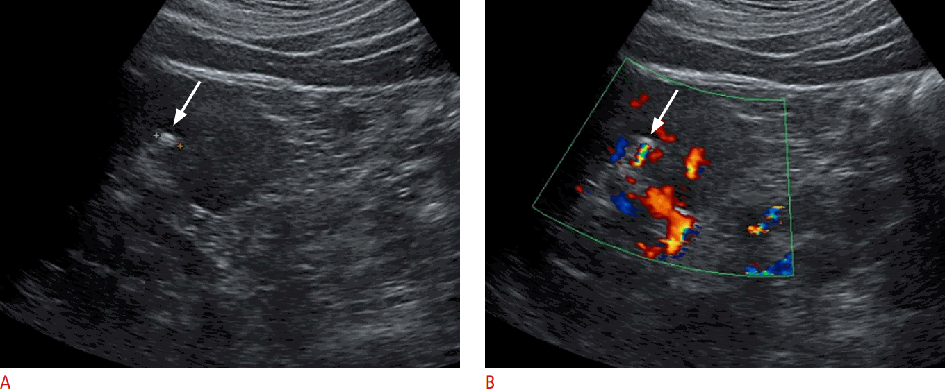
In addition, secondary findings of renal stones or hydronephrosis include the twinkling artifact and the absence of a urine jet on the affected side. The twinkling artifact is a rapid alternation of color immediately behind a stone that may be observed on color Doppler imaging, just like gallbladder stones (Fig. 4A, B) [10]. The twinkling artifact is mainly observed on rough, hyperechoic, irregular surfaces with multiple cracks, which cause a strong reflection of incident ultrasound waves. It appears as a discrete focus of alternating colors with or without an associated color comet-tail artifact [11]. The appearance of the twinkling artifact is highly dependent on the machine settings, and in order to observe it clearly, it is recommended to use a low pulse repetition frequency and high color priority [10]. Urine jets occur from the periodic contraction of the ureters and are easily visible on color Doppler imaging. Obstruction manifests as either the complete absence of the urine jet on the affected side or continuous low-level flow on the affected side, depending on the severity of the obstruction [2,6].
Clinical Considerations Regarding APN
The diagnosis of APN in adults is usually made by a combination of the typical clinical features of flank pain, elevated body temperature, and dysuria combined with abnormal urinalysis findings such as bacteriuria and/or pyuria. However, routine radiologic imaging is not required for the diagnosis and treatment of uncomplicated cases of APN in adult patients [3,12]. A radiologic evaluation may be reserved for patients with atypical symptoms, the elderly, diabetic patients, patients with congenital anomalies, and so on. US is a useful first-line tool for evaluating urinary tract infections without any radiation hazard. However, when acute bacterial pyelonephritis is suspected and an imaging work-up is required, as in complicated cases, CT is a more reasonable modality than US, because complications and possible causes (intraparenchymal or collecting system gas, a small microabscess, perinephric extension of infection, ureter stone, etc.) can be diagnosed accurately. This rationale is consistent with the American College of Radiology Appropriateness Criteria in "variant 2 of clinical condition" [13].
Ultrasonographic Evaluation of APN
The most common sonographic finding of APN is normal echogenicity. In other words, most patients with clinically suspected APN (up to 80%) have negative US results [3]. When positive findings of APN are suspected on US, they can include hypoechogenicity due to parenchymal edema and hyperechogenicity in cases of hemorrhage, swelling (Fig. 5), a perfusion defect on power Doppler images, loss of corticomedullary differentiation, wall thickening of the renal pelvis, or abscess formation (Fig. 6A, B) [3,14]. Despite the presence of several ancillary findings for the diagnosis of APN, it can be very difficult to differentiate between artifacts and true positive findings if the sonic window is poor (due to bowel gas, bony thorax, or thick subcutaneous fat tissue) or if the patient’s respiration is irregular (due to conditions such as tachypnea) (Fig. 7A, B). Thus, CT is considered to be the modality of choice for evaluating acute bacterial pyelonephritis. Another advantage of CT is that it can provide comprehensive anatomic and physiological information, thereby helping to accurately characterize both intrarenal and extrarenal pathologic conditions (Fig. 8A, B).
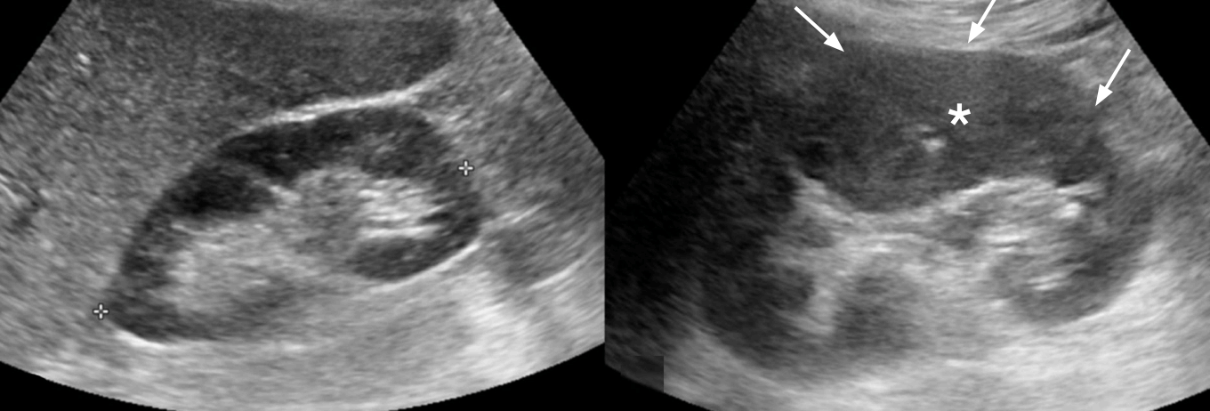
A 42-year-old woman with left flank pain and fever.
Grayscale ultrasonography shows a relatively enlarged left kidney (right panel) with increased cortical echogenicity (arrows and asterisks) compared with the right kidney (left panel). This finding may suggest parenchymal swelling due to inflammation. The urinalysis findings and clinical symptoms were also compatible with acute pyelonephritis.
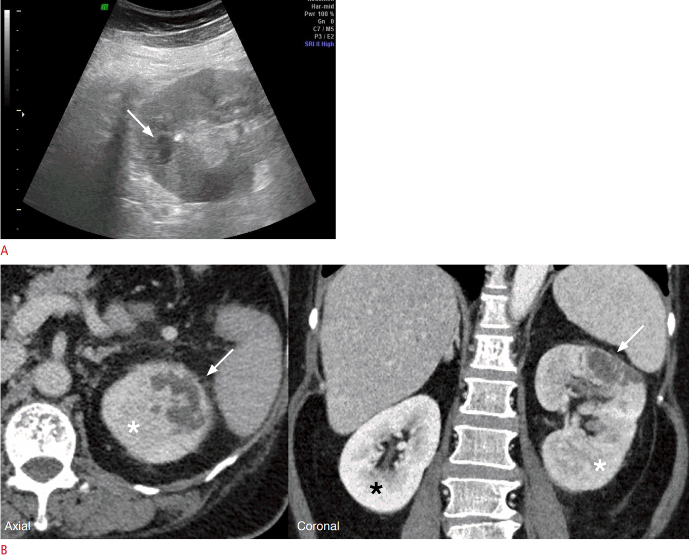
A 58-year-old woman with left flank pain and sustained fever.
During antibiotic treatment for suspected acute pyelonephritis (APN) in a private hospital, she was referred for a sustained fever. A. On a grayscale ultrasonography, the left kidney shows heterogeneously increased cortical echogenicity, and there is a small hypoechoic lesion in the upper pole (arrow). The possible cause was either a small cyst or abscess. B. A CT scan was obtained to differentiate the possibility of abscess. Portal-phase CT shows an irregularly-shaped hypodense lesion (arrows) in the upper pole of the left kidney, which is a finding highly suspicious of an abscess. Additionally, the left kidney shows decreased parenchymal enhancement (white asterisks) compared to the right kidney (black asterisk), and corticomedullary differentiation is also unclear. This finding suggests medical renal disease, such as APN.
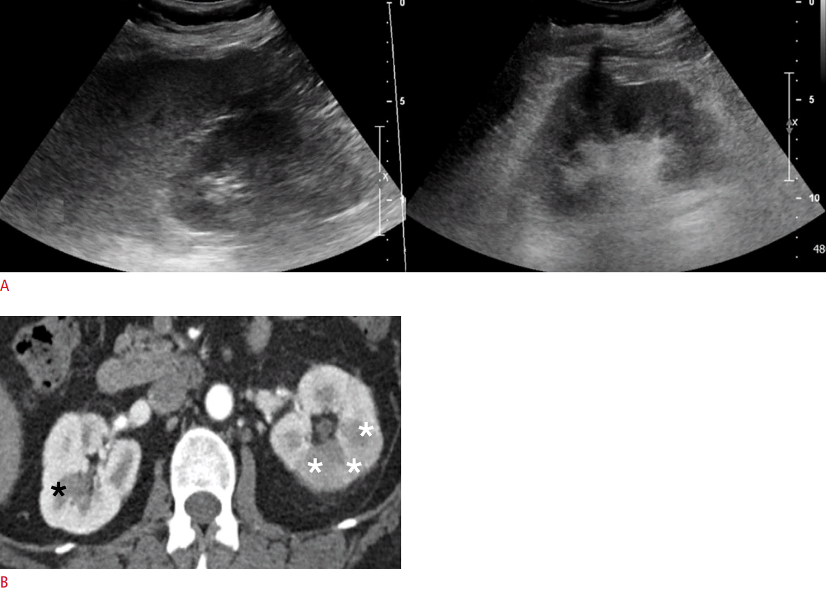
A 53-year-old woman with left flank pain.
A. On grayscale ultrasonography, it is difficult to distinguish the echogenicity difference of the right (left panel) and left kidney (right panel). B. Enhanced CT demonstrates ill-defined hypodense lesions in the left kidney (white asterisks), suggesting acute pyelonephritis. The right kidney shows normal parenchymal enhancement and corticomedullary differentiation (black asterisk). Thus, CT is considered to be the modality of choice for evaluating acute bacterial pyelonephritis.
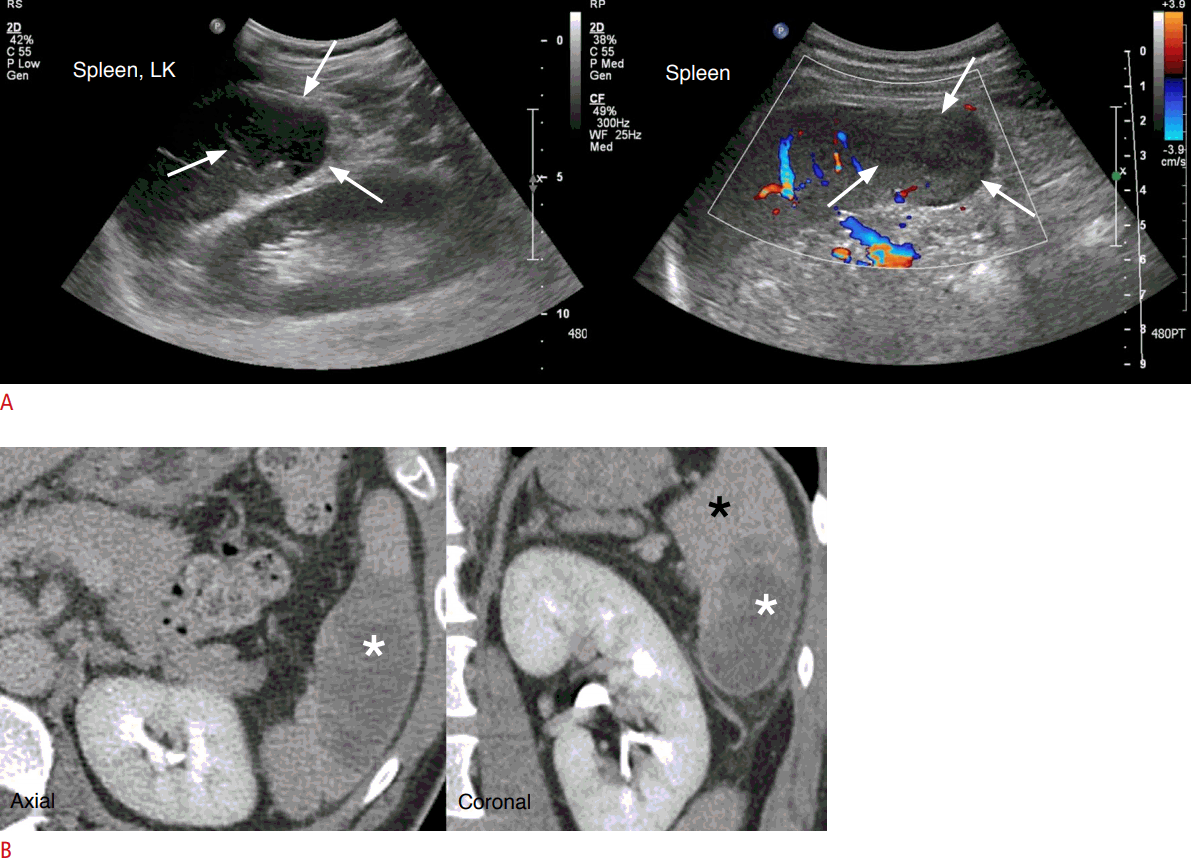
A 44-year-old man with left flank pain and leukocystosis.
A. This patient visited the emergency department due to acute left flank pain. On grayscale ultrasonography, there were no remarkable findings in the left kidney. However, hypoechoic areas are seen in the spleen (arrows). On color Doppler ultrasonography, there is an area of decreased perfusion. B. On enhanced CT, there is a hypoattenuating area in the spleen (white asterisks) compared to the normal parenchyma (black asterisk). This finding is compatible with focal splenic infarction without a demonstrable cause. Splenic infarction is a result of ischemia to the spleen but usually requires no treatment.
Mimics of Renal Colic
Unidentified bright objects
On US, tiny echogenic foci are occasionally seen in the parenchyma or hilum. These are called unidentified bright objects (UBO), and these echogenic foci frequently accompany the reverberation artifact, but posterior shadowing is absent. The possible causes of UBOs in the kidney are tiny stones, tiny cysts, small calyceal diverticulum areas with wall calcification or milk of calcium, calcified arteries, and tiny angiomyolipomas (Fig. 9).
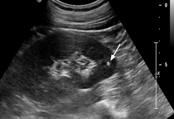
Unidentified bright objects in the kidney parenchyma.
Tiny echogenic foci (arrow) are occasionally seen in the parenchyma on ultrasonography. They are called unidentified bright objects (UBOs). These echogenic foci frequently accompany the reverberation artifact, but posterior shadowing is absent. The possible causes of UBOs in the kidney are tiny stones, tiny cysts, small calyceal diverticulum areas with wall calcification or milk of calcium, calcified arteries, and tiny angiomyolipomas. However, UBOs observed on ultrasonography may not be clearly observed on computed tomography.
Medullary nephrocalcinosis
On US, there are many causes of altered renal parenchymal echogenicity. The ultrasonographic appearance of medullary nephrocalcinosis is characterized by echogenic materials with a variable degree of acoustic shadowing in the region of the renal pyramids (Fig. 10). Depending on the degree of calcium deposition in the renal pyramid, the CT density (Hounsfield units) may also vary [15]. Medullary nephrocalcinosis occurs in various diseases as a non-specific renal manifestation. In individual cases, it is difficult to distinguish between medullary nephrocalcinosis and renal calyx calculi [16].
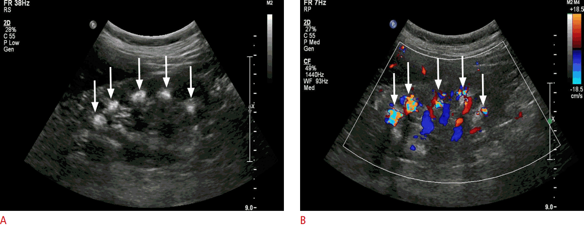
A 56-year-old woman with a medullary sponge kidney.
A. Ultrasonography shows increased echogenicity of the renal medulla (the pyramids are normally hypoechoic to the cortex). This appearance is typical of medullary nephrocalcinosis (arrows). B. Color Doppler ultrasonography shows twinkling artifacts (arrows) at the renal medulla.
Extrarenal pelvis
Extrarenal pelvis refers to the presence of the renal pelvis outside the confines of the renal hilum. It is a normal variant that is found in ~10% of the population. The renal pelvis is formed by all the major calyces. An extrarenal pelvis usually appears as a dilated pelvocaliceal system giving a false impression of obstructive hydronephrosis. Subsequent investigation with CT usually clarifies the false interpretation on US (Fig. 11).

Extrarenal pelvis.
A. Grayscale ultrasonography of the right kidney shows dilatation of the renal pelvis (arrows). A beginner could mistakenly think that mild hydronephrosis was present. B, C. A prominent extrarenal pelvis (asterisks) is a well-known normal variation that mimics hydronephrosis on excretory axial CT (B) and intravenous pyelography (C). Note that neither calyceal blunting on an intravenous pyelogram nor contrast excretion delay of the right kidney is present.
Parapelvic cysts
Parapelvic cysts of the kidneys are simple renal cysts that are adjacent to the renal pelvis or the renal sinus. Parapelvic cysts do not communicate with the collecting system and are probably lymphatic in origin or develop from embryonic remnants [17]. Most are asymptomatic, and treatment is rarely necessary unless they cause hematuria, hypertension, hydronephrosis, or become infected [17]. Parapelvic cysts demonstrate stretching and compression of the calyces on intravenous pyelograms (Fig. 12A), similar to the appearance of marked renal sinus lipomatosis. On US, they have the typical appearance of centrally located cysts, but may be mistaken for hydronephrosis (Fig. 12B). However, excretory CT shows multiple cystic lesions both in the renal hilum, with intervening normal renal pelvis areas, and in calyces filled with a contrast agent (Fig. 12C).

Parapelvic cysts.
A. On an intravenous pyelogram, stretching and compression of the calyces (arrows) are noted in both kidneys. B. Grayscale ultrasonography shows dilatation of the renal pelvis and calyces (asterisks) in both kidneys mimicking bilateral hydronephrosis. C. Excretory coronal CT shows multiple cystic lesions (asterisks) in the bilateral renal hila with intervening normal renal pelvis and calyces.
Conclusion
Although US has several limitations in adults with acute flank pain, it is a useful modality to diagnose stones and to confirm the occurrence of complications of APN, so it is important to understand these characteristic findings and other diseases that mimic them. In addition, other imaging modalities such as CT can be recommended if the clinical or radiological diagnosis is ambiguous.
Notes
No potential conflict of interest relevant to this article was reported.

