Ultrasonography for nerve compression syndromes of the upper extremity
-
Soo-Jung Choi
 , Jae Hong Ahn
 , Dae Shik Ryu
 , Chae Hoon Kang
 , Seung Mun Jung
 , Man Soo Park
 , Dong-Rock Shin
- Received December 11, 2014 Revised January 12, 2015 Accepted January 13, 2015
- ABSTRACT
-
Nerve compression syndromes commonly involve the nerves in the upper extremity. High-resolution ultrasonography (US) can satisfactorily assess these nerves and may detect the morphological changes of the nerves. US can also reveal the causes of nerve compression when structural abnormalities or space-occupying lesions are present. The most common US finding of compression neuropathy is nerve swelling proximal to the compression site. This article reviews the normal anatomic location and US appearances of the median, ulnar, and radial nerves. Common nerve compression syndromes in the upper extremity and their US findings are also reviewed.
- Introduction
- Introduction
The development of high-frequency linear array transducers has led to an improvement in the resolution of ultrasonography (US), and thus, US can nowadays satisfactorily depict most of the peripheral nerves. Therefore, US has been widely used, along with magnetic resonance imaging (MRI) and a nerve conduction study, in cases of common nerve compression syndromes or other neuropathic conditions including trauma. Although MRI is very accurate and superior for imaging soft tissues and nerves, it is expensive, takes more time, and may have artifacts. More importantly, MRI may not be practical when a number of nerves need to be examined over the long course [1]. Although US has a few limitations like a small field of view, limited penetration, and an anisotropic effect when imaging nerves [2], major nerves in the upper extremity can usually be visualized without much difficulty. US of peripheral nerves requires the use of transducers with high insonation frequencies (7-12 MHz or more). In this article, we review the anatomy and US features of the main nerves of the upper extremity and evaluate the clinical and US features of common nerve compression syndromes of the upper extremity.
- Median Nerve
- Median Nerve
- Pronator Teres Syndrome
- Pronator Teres Syndrome
Median nerve entrapment can occur around the elbow joint. The most common site of median nerve compression at the elbow level is between the two heads of the pronator teres muscle [3]. The patients having median nerve compression at this level may have pain and numbness in the volar side of the elbow and forearm. The hand symptoms may be similar to those in the case of carpal tunnel syndrome (CTS). This is called pronator teres syndrome. The causes of pronator teres syndrome may relate to trauma with hematoma, pronator teres muscle hypertrophy, or congenital abnormalities [4]. The demonstration of the causes of the pronator teres syndrome by US may be possible if there is a mass or hematoma causing compression of the median nerve. However, in the absence of such findings, it may not be easy to make a diagnosis or to find the cause of nerve compression with US, since the fibrous bands or scar tissue may be too small to visualize with US [5]. MRI can demonstrate the denervation muscle signal changes and determine the level of nerve compression [6].- AIN Syndrome (Kiloh-Nevin Syndrome)
- AIN Syndrome (Kiloh-Nevin Syndrome)
AIN syndrome is caused by entrapment or compression of the AIN. The AIN innervates muscles in the deep anterior compartment of the forearm: FPL, radial-side FDP, and PQ. Lesions of the AIN can result in a variety of clinical symptoms, depending on the location and the degree of nerve damage [7]. Typically, the patients have motor disturbance in the function of the affected muscles and weakness of the thumb, index finger, and occasionally the middle finger. Therefore, patients with AIN syndrome have the characteristic circle sign; that is, these patients cannot form an “O” shape with their index finger and thumb due to the lack of innervation of the FPL and FDP of the index finger [4]. Some patients may have isolated weakness of the thumb because of the isolated involvement of the particular fascicle that innervates the FPL [8]. The causes of AIN syndrome can be external compression with hematoma or mass or direct nerve trauma [5]. However, some patients without direct trauma or demonstrable causes may have AIN syndrome. The AIN is usually compressed by fibrous bands, most commonly originating from the deep head of the pronator teres, flexor digitorum superficialis, and brachialis fascia [7]. Although US is usually limited for the evaluation of AIN syndrome because of the small size of the nerve and relatively deep location, US can identify the anterior interosseous artery at the volar aspect of the interosseous membrane by using color or power Doppler. The identification of the anterior interosseous artery helps in the evaluation of the lesion along the course of the AIN adjacent to the anterior interosseous artery [5]. In some cases, US may identify the small volume and denervation echo changes of the denervated muscles (Fig. 2).- Carpal Tunnel Syndrome
- Carpal Tunnel Syndrome
CTS is the most common nerve compression syndrome in the upper extremity. The causes of CTS are diverse depending on the origin: tenosynovitis of flexor tendons (keyboard operator/inflammatory arthritis), space-occupying lesions such as ganglions or tumors (Fig. 3), nerve swelling related to thyroid disease or pregnancy, amyloid deposition, post-traumatic fibrosis, excess fat, anatomic variation such as a persistent median artery with or without a bifid median nerve (Fig. 4), and anomalous muscle [9]. In most cases, the diagnosis of CTS is usually made on the basis of typical clinical signs and symptoms, and an electrodiagnostic study is performed to confirm the diagnosis [9]. US has been used for patients with CTS as an additional diagnostic tool, since US can depict the morphological changes and size abnormalities of the median nerve and demonstrate the cause of nerve compression [10-15]. On US, the normal median nerve is round in the distal forearm, oval at the level of the pisiform, and flat at the level of the hook of hamate (Fig. 5) [16]. The most common US finding of CTS is the enlargement of the median nerve, and this is usually most prominent at the scaphoid-pisiform level. There have been many studies aiming to provide the most appropriate cutoff value of the median nerve cross-sectional area for the diagnosis of CTS [10-14,17]. By using the ellipse formula, researchers have suggested a cutoff value between 9 mm2 and 10 mm2 at the scaphoid-pisiform level as the diagnostic criteria of CTS with acceptable sensitivity and specificity [13,14,17,18]. According to Buchberger et al. [17,19], four characteristic findings can be found in patients with CTS: (1) a significant increase in the cross-sectional area of the median nerve at the level of the pisiform and, to a lesser degree, at the level of the hamate; (2) a significant increase in the cross-sectional area of the median nerve at the level of the pisiform as compared to that at the level of the distal radius (swelling ratio); (3) a significant increase in the flattening ratio at the level of the hook of hamate; and (4) significant palmar bowing of the flexor retinaculum (palmar displacement of the flexor retinaculum of more than 4 mm from the line between the hook of hamate and the tubercle of the trapezium). US could also reveal the hypoechoic change of the median nerve and the altered fascicular pattern caused by neural edema (Fig. 6). In addition, US is useful for the postoperative evaluation of CTS [20,21]. According to the studies by Ozturk et al. [20] and Apfelberg and Larson [21], US could reveal the decrease in the cross-sectional area of the median nerve after transverse carpal ligament release. US may demonstrate the causes of recurrent CTS after surgery (Fig. 7).
The median nerve arises from the medial and lateral cord of the brachial plexus and runs down along the medial side of the distal arm with the brachial artery and vein. It passes through the antecubital fossa deeply relative to the biceps aponeurosis (also called the lacertus fibrosus) and anterior to the brachialis muscle. It is located between the biceps brachii and the brachialis muscle at the elbow level. The nerve lies between the humeral (superficial) and ulnar (deep) heads of the pronator teres muscle at the level of the elbow joint [3]. Transverse US with a high-resolution linear array transducer (5-17 MHz) can demonstrate the normal median nerve between the two heads of the pronator teres muscle (Fig. 1). The anterior interosseous nerve (AIN) arises from the median nerve at the level of the humeral head of the pronator teres muscle and travels along the anterior aspect of the interosseous membrane of the forearm between the flexor pollicis longus (FPL) and the flexor digitorum profundus (FDP) muscles. The AIN is a purely motor nerve and supplies the pronator quadratus (PQ) muscle, FPL muscle, and radial-side FDP muscle. At US, the identification of the normal AIN may be difficult because of the deep location and small size of the nerve. After exiting the pronator teres muscle, the main median nerve enters the anterior compartment of the forearm by passing beneath the fibrous arch of the heads of the flexor digitorum superficialis muscle and runs down to the wrist level to enter the carpal tunnel.
- Ulnar Nerve
- Ulnar Nerve
- Cubital Tunnel Syndrome
- Cubital Tunnel Syndrome
Cubital tunnel syndrome is the second most common nerve compression syndrome in the upper extremity [4]. Cubital tunnel syndrome is caused by external compression or injury of the ulnar nerve within the cubital tunnel. The possible causes of this syndrome include bone fragments, loose bodies or the spur arising from the elbow joints, space-occupying lesions such as tumors, ganglia, hematomas, synovitis from the elbow joint, regional ligamentous inflammation, accessory muscle (anconeus epitrochlearis), and dislocation of the nerve. Patients with cubital tunnel syndrome may present medial elbow pain, numbness in the ring and little fingers, and weakness in the intrinsic hand muscles. Patients with chronic cubital tunnel syndrome may show a claw hand deformity due to muscle volume loss in the first interosseous space and hypothenar eminence and semi-flexion deformity of the ring and the little fingers [4]. The ultrasonographic appearance of cubital tunnel syndrome is the enlargement of the ulnar nerve at the level of the cubital tunnel, similar to CTS (Fig. 11). There have been several reports aiming to provide the cutoff value of the ulnar nerve cross-sectional area for the diagnosis of cubital tunnel syndrome [22-24]. According to these previous studies, the cutoff point of 10 mm2 or higher for the cross-sectional area led to a sensitivity and specificity of more than 88% for the diagnosis of cubital tunnel syndrome [22,23]. Thoirs et al. [24] suggested a cutoff value for the crosssectional area of the asymptomatic ulnar nerve of 9 mm2, derived as the upper limit of the 95% confidence interval. An additional study used a ratio of 1.5:1, comparing the ulnar nerve area at the level of the cubital tunnel with that proximal to the cubital tunnel, and the 8.3-mm2 cross-sectional area of the ulnar nerve at the epicondyle level showed 100% sensitivity in the diagnosis of cubital tunnel syndrome [25]. The ulnar nerve in patients with cubital tunnel syndrome is usually hypoechoic in US because of neural edema. In cases of chronic cubital tunnel syndrome, US may identify the echogenic FCU and ulnar-side FDP muscles with small volume because of the muscle hypotrophy caused by chronic denervation.- Guyon’s Canal Syndrome
- Guyon’s Canal Syndrome
Guyon’s canal is an oblique fibro-osseous tunnel with a length of about 4 cm between the hook of hamate and the pisiform. The borders of Guyon’s canal vary along its entire course and are not distinct [26]. The roof of the canal consists of the palmar aponeurosis, palmaris brevis, and hypothenar fibroadipose tissue; the floor is made up of the FDP tendons, transverse carpal ligament, pisohamate ligament, pisometacarpal ligament, and opponens digiti minimi; the medial wall is composed of the FCU tendon, the pisiform, and the abductor digiti minimi; and the lateral wall is formed by the extrinsic flexor tendons, the hook of hamate, and the transverse carpal ligament [27-29]. The contents of Guyon’s canal are the ulnar artery, nerve, communicating vein, and loose fibrofatty tissue. Guyon’s canal syndrome is a compressive neuropathy of the ulnar nerve at the level of Guyon’s canal. One cause of Guyon’s canal syndrome is related to trauma. Direct trauma may cause a nerve injury related to the compression between the external force and the hook of hamate [5]. Repetitive trauma to the hypothenar area by the handlebar can happen in cyclists and can cause Guyon’s canal syndrome. This is called cyclist’s palsy. “Hypothenar hammer syndrome” results from direct trauma to the ulnar artery between the hook of hamate and the palmaris brevis in those using jackhammers or people using vibratory tools occupationally [26]. Repetitive trauma to the ulnar artery can cause thrombosis and occlusion of the ulnar artery and resultant ulnar nerve compression (Fig. 12). Fracture of the hook of hamate or other carpal bones can also cause Guyon’s canal syndrome. A ganglion is one of the most common causes of Guyon’s canal syndrome [26], and other space-occupying lesions including tumorous conditions can cause ulnar nerve compression as well. Other causes of Guyon’s canal syndrome are anomalous muscles, abnormally thickened ligaments, and an anomalous course of the ulnar nerve [26]. US can identify the cause of Guyon’s canal syndrome and the morphologic change of the ulnar nerve (Fig. 13).
The ulnar nerve arises from the medial cord of the brachial plexus and runs down along the m0 level, the nerve penetrates the medial intermuscular septum to enter the posterior compartment and descends along the anterior and medial aspects of the triceps muscle. At the distal arm level, the ulnar nerve passes posterior to the medial humeral epicondyle and finally enters the cubital tunnel [3]. The cubital tunnel is a fibro-osseous channel formed by the olecranon process laterally, the posterior cortex of the medial epicondyle medially, the elbow joint capsule and posterior bundle of the medial collateral ligament anteriorly, and the cubital tunnel retinaculum (Osborne ligament) posteriorly [3]. The nerve exits the distal aspect of the cubital tunnel to enter the medial aspect of the forearm between the superficial and deep heads of the flexor carpi ulnaris (FCU) muscle. At the elbow level, the ulnar nerve gives off motor branches to the FCU and ulnar-side FDP muscles [4]. At the wrist level, it passes through another fibro-osseous canal called Guyon’s canal and branches into superficial sensory and deep motor nerves within the canal. High-resolution US can demonstrate the normal ulnar nerve at the level of the cubital tunnel and the wrist. The ulnar nerve is easy to identify in the cubital tunnel between the medial humeral epicondyle and the olecranon process with transverse US (Fig. 8). The elbow should be extended during the initial evaluation since the ulnar nerve may be dislocated or displaced anteriorly during elbow flexion (Fig. 9) [5]. According to the study of Ozturk et al. [20] on the ultrasonographic appearance of the normal ulnar nerve in the cubital tunnel in 212 elbows, 77.8% of the cases showed the ulnar nerve with one fascicle, and the rest showed two or three fascicles at the level of the cubital tunnel. The size and morphology of the ulnar nerve can differ with the elbow flexion position and the extension position [20,21]. During elbow flexion, 31.6% of ulnar nerves showed anterior displacement, and the volume of the cubital tunnel and the cross-sectional area of the ulnar nerve decreased [20]. At the level of Guyon’s canal, the hyperechoic cortex of the pisiform can be used as a landmark to find the ulnar nerve. The normal fibrillar pattern of the ulnar nerve is well demonstrated in Guyon’s canal with US, and bifurcation of the ulnar nerve is identified (Fig. 10).
- Radial Nerve
- Radial Nerve
- Radial Tunnel Syndrome and PIN Syndrome
- Radial Tunnel Syndrome and PIN Syndrome
PIN syndrome is a compressive neuropathy of the deep branch of the radial nerve in the region of the supinator muscle [33]. It can have various etiologies. Most commonly, repetitive overuse of the forearm (repetitive pronation-supination or flexion-extension) can cause a radial nerve injury in this region [34]. This is common to athletes (tennis players). The thickened or hypertrophied anatomic structures can entrap the nerve, such as a thickened proximal tendinous edge of the supinator muscle (arcade of Frohse), thickened leading edge of the extensor carpi radialis brevis muscle, and distal ligamentous margin of the supinator muscle [30]. The arcade of Frohse is the most common area of compression. Other causes are space-occupying lesions (such as tumor/ganglion/bicipitoradial bursitis), prominent recurrent radial vessels, fascial band of the radial head, and systemic diseases such as diabetes, rheumatoid arthritis, polyarteritis, neuralgic amyotrophy, chronic hypoperfusion, and postsystemic illness angioneuropathy [35]. Clinical manifestation of the PIN syndrome varies depending on the level of nerve compression and the affected nerve fibers [34]. Compression of the deep branch of the radial nerve mostly causes painless palsy of the extensor muscles of the forearm. However, tenderness and pain at the lateral aspect of the proximal forearm mimicking lateral epicondylitis can be clinically presented without motor weakness in some patients. This is referred to as the “radial tunnel syndrome” (Fig. 18) [3]. The most commonly observed US finding of PIN syndrome is the enlargement of the PIN at the proximal portion of the compression site [33-37]. According to the study of Djurdjevic et al. [33] on high-resolution US findings in 13 patients with PIN syndrome, US was able to reveal the edema and caliber change of the PIN in all patients. Nine of these 13 patients underwent surgery in this study and intraoperative exploration showed PIN compression by the tight tendinous arcade of Frohse in all of the patients. The researchers also suggested a 15-mm cutoff value of the PIN diameter for the diagnostic criteria of PIN syndrome by comparison with a normal control group. US can also show the presence of hyperemia of the nerve on color Doppler imaging [37]. Rarely, space-occupying lesions causing PIN compression can be found with US. In addition, US may reveal an echo difference of the dorsal extensor muscles caused by denervation, as compared to the contralateral side.- Wartenberg’s Syndrome
- Wartenberg’s Syndrome
Wartenberg’s syndrome is an entrapment neuropathy of the superficial branch of the radial nerve at the level of the distal forearm and wrist [32]. The patients present with pain, numbness, and paresthesia in the radial-side wrist and thumb. Therefore, de Quervain’s disease and intersection syndrome should be differentiated. The superficial branch of the radial nerve is easily injured by trauma. The nerve can be injured during the placement of fixator pins due to Colles fractures. The superficial branch of the radial nerve injury by penetrating trauma, and cephalic vein cannulation are also reported [38]. Some physical activities require forearm pronation with simultaneous flexion and ulnar deviation of the hand predisposed to superficial radial nerve entrapment between the brachioradialis muscle and the extensor carpi radialis longus muscle [32]. Wartenberg’s syndrome is commonly associated with de Quervain’s disease, although the reason for this is not yet known [31]. US is helpful in distinguishing Wartenberg's syndrome from de Quervain's disease or arthritis of the trapeziometacarpal joint [32]. High-resolution US may depict the abnormal changes of the superficial branch of the radial nerve [5,31,32,33]. US in patients with Wartenberg’s syndrome can show secondary nerve compression from the adjacent scar tissue or post-traumatic/postoperative changes of the soft tissue. These changes could appear as abnormal heterogeneous hypoechoic tissue [5].
The radial nerve is the terminal branch of the posterior cord of the brachial plexus. At the arm level, it obliquely runs posterior to the humerus in the spiral groove. The radial nerve is closely attached to the posterior cortex of the humerus in the spiral groove, at this level (Fig. 14). At the level of the distal arm, the radial nerve pierces the intermuscular septum to enter the anterior aspect of the humeral lateral condyle. Anterior to the lateral epicondyle, the radial nerve divides into a superficial sensory branch and a deep motor branch [4]. The superficial sensory branch runs in the forearm along the deep surface of the brachioradialis muscle along with the radial artery. After bifurcation of the radial nerve, the deep motor branch enters the radial tunnel [3]. The radial tunnel extends from the level of the radiocapitellar joint to the level of the proximal aspect of the supinator muscle. The radial tunnel is bounded by the joint capsule posteriorly, the brachialis muscle and biceps tendon medially, and the brachioradialis muscle and the extensor carpi radialis brevis and longus muscles laterally. At the distal aspect of the radial tunnel, the deep branch pierces the supinator muscle and passes through the supinator muscle to enter the posterior compartment of the forearm (Fig. 15). Thereafter, the deep motor branch of the radial nerve leaves the supinator muscle; this branch is referred to as the posterior interosseous nerve (PIN). At the level of the upper arm, the radial nerve gives off branches to innervate the triceps and anconeus muscles. At the level of the radial tunnel, the radial nerve gives off branches to innervate the brachioradialis, extensor carpi radialis longus and brevis, and supinator muscles, in this order. After exiting the supinator muscle, the PIN innervates the dorsally located extensor muscles (the extensor digitorum, extensor carpi ulnaris, extensor digiti minimi, abductor pollicis longus, extensor pollicis brevis, extensor pollicis longus, and extensor indicis muscles) [30]. US can identify the normal radial nerve in the spiral groove, over the posterior aspect of the humerus (Fig. 14). The radial nerve in the radial tunnel is also easy to identify in the anterior elbow with transverse US. The fascicular pattern of the radial nerve can be seen in the fascial plane between the brachioradialis (laterally) and brachialis (deep medially) muscles [5]. When the radial nerve is traced down, the bifurcation of the radial nerve is seen. More distally, the deep motor branch can be followed with transverse US as it enters the supinator muscle. An oblique scan at the level of the supinator muscle can demonstrate the deep motor branch passing through the supinator muscle and continuing as the PIN (Fig. 16). The superficial branch of the radial nerve runs down along the deep portion of the brachioradialis and becomes superficial at about 9 cm proximal to the styloid process of the radius [31]. More distally, it divides into three separate branches. US is able to identify the superficial branch of the radial nerve crossing the first extensor compartment of the wrist (Fig. 17). At the level of the distal forearm, the superficial radial nerve lies close to the cephalic vein [32].
- Conclusion
- Conclusion
High-resolution US is a useful method of examination for the evaluation of peripheral nerves, particularly the median, ulnar, and radial nerves of the upper extremity. However, the quality of US may differ depending on the examiner’s knowledge. The anatomic location and normal ultrasonographic appearances of the peripheral nerves should be noted when the US exam is performed. Radiologists should also inform themselves about common nerve compression syndromes in the upper extremity and their causes to make an accurate assessment of the disease.
- NOTES
- NOTES
No potential conflict of interest relevant to this article was reported.
Fig. 1.
Schematic representation and transverse sonograms of the median nerve at the elbow level.
A. The schematic representation shows the relationship between the pronator teres muscle (shaded in blue color) and the median nerve (colored in yellow). The median nerve enters between the humeral (superficial) and ulnar (deep) heads of the pronator teres muscle at the level of the elbow joint. Dashed arrows inserted in the schematic indicate the corresponding levels of the sonograms. B. At the level of the distal arm, the transverse sonogram shows the median nerve (arrow) running along the brachial artery and vein. C. At the level of brachial artery bifurcation into radial and ulnar artery, the median nerve (arrow) follows the ulnar artery and the accompanying vein. D, E. At the level of the pronator teres muscle, the median nerve (arrows) is found between the two heads of the pronator teres muscle. PT-h, humeral head of the pronator teres muscle; PT-u, ulnar head of the pronator teres muscle. F. After exiting the pronator teres muscle, the median nerve enters the anterior compartment of the forearm (arrows). Although the anterior interosseous nerve arises from the median nerve at this level, the transverse sonogram only shows the color Doppler signal from the anterior interosseous artery from the anterior aspect of the volar side interosseous membrane. FDP, flexor digitorum profundus; FPL, flexor pollicis longus.

Fig. 2.
A 43-year-old male patient with anterior interosseous nerve syndrome (Kiloh-Nevin syndrome).
A. The transverse sonogram at the level of the mid-forearm shows prominently increased echogenicity in the flexor pollicis longus (FPL) and radial-side flexor digitorum profundus (FDP-r) muscles. Note the normal echogenicity of the superficially located muscles. FDS, flexor digitorum superficialis muscle. No space-occupying lesion was found along the course of the anterior interosseous nerve and vessels (arrows). B. The transverse sonogram at the level of the distal forearm reveals the echogenic pronator quadratus (PQ) muscle. C. The transverse sonogram at the same level as that of B in the contralateral forearm shows normal echogenicity of the PQ muscle. D, E. Axial fat-suppressed T2-weighted images of the same patient reveal signal changes in the FPL, FDP-r (in D), and PQ muscles (in E) innervated by the anterior interosseous nerve.
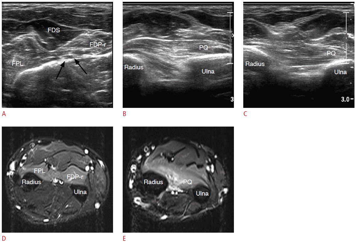
Fig. 3.
A 37-year-old female patient with carpal tunnel syndrome.
A, B. Longitudinal (A) and transverse (B) sonograms at the level of the wrist, just distal to the carpal tunnel show a mass-like enlargement of the median nerve (arrows). The nerve fascicles inside the median nerve are also thickened and look like electronic cables or spaghetti noodles. C. T1-weighted axial magnetic resonance image shows an enlarged median nerve with a characteristic T1 high-signal intensity coaxial cable appearance (arrows). The mass was surgically removed and proved to be the fibrolipomatous hamartoma of the median nerve (Courtesy of MH Lee, Seoul Asan Medical Center).
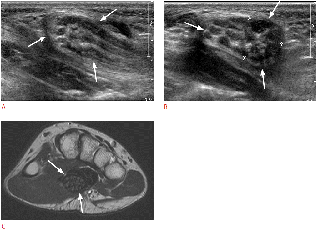
Fig. 4.
A 46-year-old female patient with incidentally found bifid median nerve (MN) and persistent median artery.
A, B. Ultrasonography (A) and magnetic resonance imaging (B) were conducted for the evaluation of the soft tissue mass in her hand (data not shown). At the level of the wrist (proximal to carpal tunnel), the transverse sonogram (A) and the axial fat-suppressed T2-weighted magnetic resonance image (B) show the median nerve having two separate bundles (arrows) and persistent median artery (arrowheads) in between. This patient did not present any neurologic symptoms.

Fig. 5.
Transverse sonograms of the normal median nerve (MN) at the level of the carpal tunnel.
A. At the level of the proximal carpal tunnel, scaphoid (S) and pisiform (P) are landmarks to indicate the proximal carpal tunnel. Note the transverse carpal ligament (arrows) forming the roof of the carpal tunnel. The ulnar artery (UA) and nerve (UN) are seen right next to the pisiform, in Guyon’s canal. The MN is seen as a hypoechoic oval structure, as compared to the adjacent tendons. The normal fascicular pattern of the nerve is well seen. FCR, flexor carpi radialis tendon; FDP, flexor digitorum profundus; FPL, flexor pollicis longus; FDS, flexor digitorum superficialis. B. At the level of the distal carpal tunnel, the trapezium (T) and the hook of hamate (H) are the landmarks of the distal carpal tunnel. Flexor retinaculum looks flat as compared to that in the proximal carpal tunnel (arrows). The MN looks hypoechoic due to anisotropy, and hyperechoic epineural fat is seen around this nerve.

Fig. 6.
A 68-year-old female patient with carpal tunnel syndrome.
A. The transverse sonogram at the level of the distal radioulnar joint shows enlargement of the median nerve. The cross-sectional area (CSA) of the median nerve is 15.4 mm2. B. At the level of the proximal carpal tunnel, CSA of the median nerve is 30.5 mm2. The normal fascicular pattern of the median nerve is not seen because of the neural edema. Flexor tendons are hypoechoic due to anisotropy. S, scaphoid; P, pisiform. C. At the level of the distal carpal tunnel, CSA of the median nerve is 22.2 mm2, showing that the nerve is still enlarged but smaller than that in the proximal carpal tunnel. The median nerve looks flat and linear due to entrapment. The normal epineural fat around the median nerve is not seen. T, trapezium; H, hook of hamate. D. Longitudinal sonogram at the midline wrist and palm shows a wavy contour of the median nerve caused by proximal swelling (arrowheads) and distal entrapment (arrows).
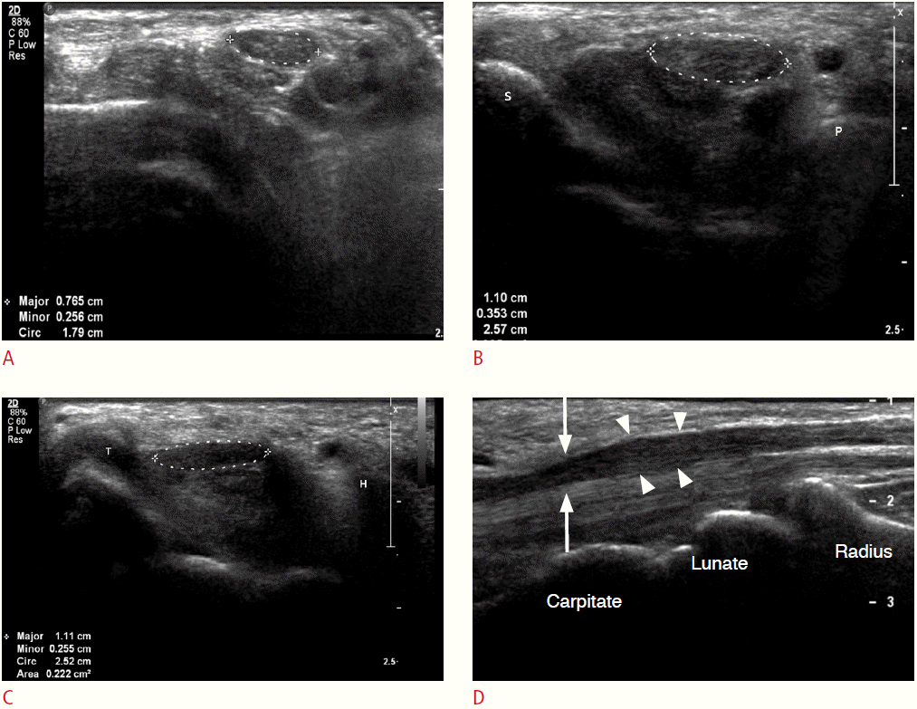
Fig. 7.
A 59-year-old female patient with recurrent carpal tunnel syndrome.
A. The transverse sonogram shows turbid fluid collection around the flexor tendons (arrows), suggesting flexor tenosynovitis. B. Axial T2-weighted magnetic resonance image also shows fluid collection around the flexor tendons (arrows). No retinacular regrowth or fibrosis is noted. The cause of the recurrent carpal tunnel syndrome in this patient was considered to be flexor tenosynovitis.

Fig. 8.
The schematic representation and transverse sonograms of the normal ulnar nerve in the cubital tunnel and the proximal forearm.
A. The schematic shows the posterior aspect of the elbow. Dashed arrows inserted in the schematic indicate the corresponding levels of the sonograms. FCU, flexor carpi ulnaris muscle. B. The transverse sonogram at the level of the cubital tunnel shows the normal ulnar nerve (arrow), posterior to the medial epicondyle (ME). Note the cubital tunnel retinaculum (arrowheads) from the ME to the olecranon process (O). C. More distally, the ulnar nerve (arrows) is exiting the cubital tunnel. The cubital tunnel retinaculum (arrowheads) is seen between two muscle bellies of the FCU. D. After exiting the cubital tunnel, the ulnar nerve (arrow) is seen between two muscle bellies of FCU. E. The transverse sonogram at the level of the ulnar-side proximal arm shows normal ulnar nerve (arrow) embedded in the FCU muscles.
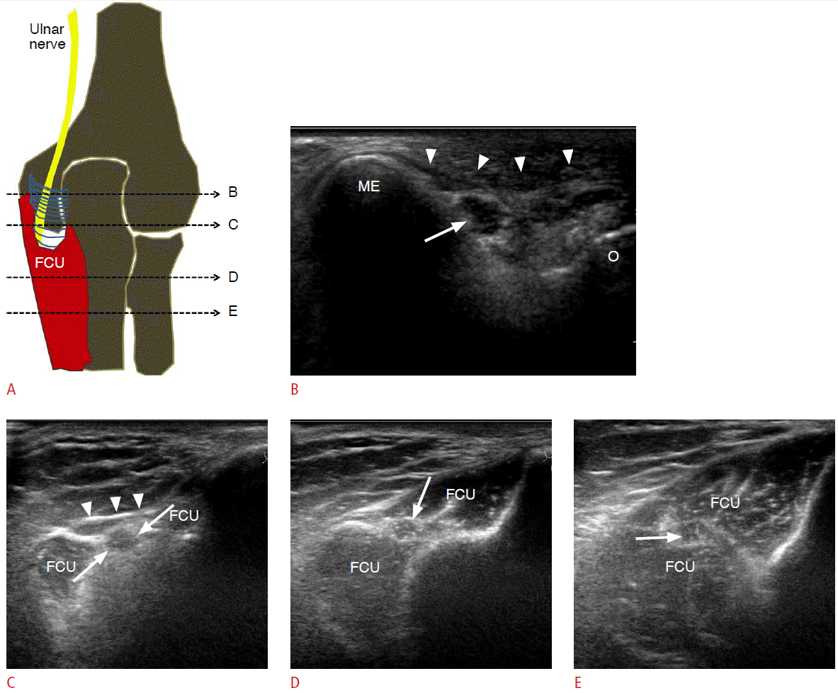
Fig. 9.
The ulnar nerve location for different elbow positions.
A. The transverse sonogram during elbow extension shows the ulnar nerve (arrow) in the cubital tunnel. Note the cubital tunnel retinaculum (arrowheads). ME, medial epicondyle. B. The transverse sonogram during elbow flexion in the same patient shows anterior displacement of the ulnar nerve (arrow). However, the ulnar nerve is still located behind the ME; no subluxation or dislocation is observed.

Fig. 10.
Transverse sonograms of the normal ulnar nerve in Guyon’s canal.
A. The transverse sonogram at the level of the pisiform (P) shows the normal ulnar nerve (arrow) between the pisiform (P) and the ulnar artery (arrowhead) in Guyon’s canal. B. More distally, the sonogram at the level of the hook of hamate (H) shows two branches of the ulnar nerve (superficial sensory and deep motor branch) (arrows). Note the flexor retinaculum (arrowheads) attaching to the hook of hamate.
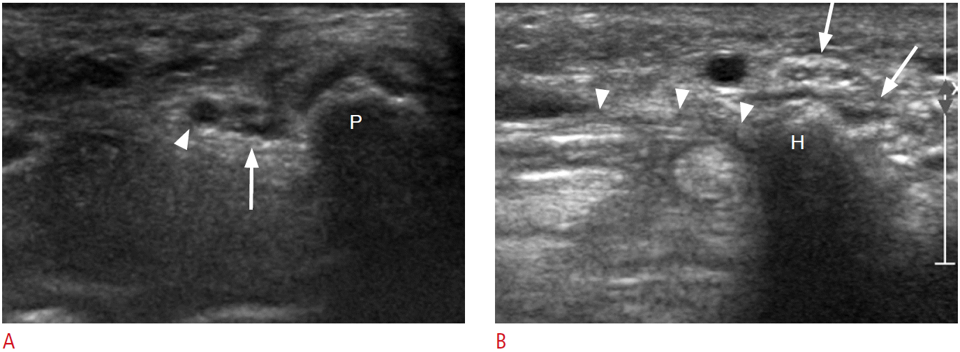
Fig. 11.
A 64-year-old male patient with cubital tunnel syndrome.
A. The elbow anteroposterior view shows severe osteoarthritis in the elbow joint. Note multiple spur changes and loose bodies in the medial side of the joint. B, C. Transverse sonograms at the level of the cubital tunnel show prominent enlargement of the ulnar nerve (arrows). The cross-sectional area of the ulnar nerve is 30.2 mm2. The ulnar nerve looks hypoechoic due to neural edema. The normal fascicular pattern is lost. D. The longitudinal sonogram of the ulnar nerve shows segmental enlargement and swelling at the level of the elbow joint. The left side of the picture shows the distal part of the upper extremity. E. An operative photograph of the same patient shows the prominently swollen ulnar nerve (arrows). Cubital tunnel release was performed in this patient.
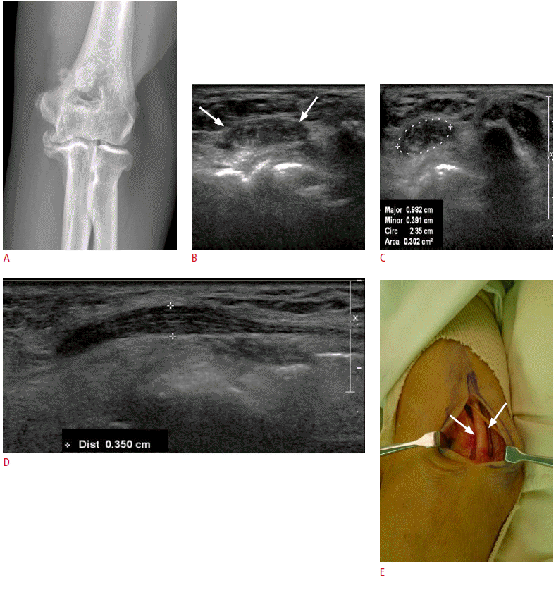
Fig. 12.
A 54-year-old male patient with Guyon’s canal syndrome.
The patient had fallen to the ground and caught himself with his hand 3 months previously, and suffered from numbness and pain in the ulnar side of his hand for 1 month. A. The transverse sonogram at the level of the wrist shows a soft tissue mass (black arrows) adjacent to the superficial branch of the ulnar nerve (white arrow). B. The longitudinal sonogram reveals the relationship between the ulnar artery (UA) and the mass (arrows). The ultrasonographic diagnosis was UA thrombosis and resultant ulnar nerve compression. C. An operative photograph shows thrombosis in the UA (arrows). The ulnar nerve was tightly adhered to the thrombosis (data not shown).

Fig. 13.
A 67-year-old female patient with Guyon’s canal syndrome.
A. The transverse sonogram at the level of the wrist (just proximal to Guyon’s canal) shows the normal ulnar nerve with a fascicular pattern (arrows). UA, ulnar artery. B. The transverse sonogram at the level of the pisiform (P) shows an intraneural hypoechoic lesion in the ulnar nerve (arrows). C. The longitudinal sonogram at the level of Guyon’s canal reveals a neuroma (arrows) and a preserved nerve fascicle (arrowheads) within the ulnar nerve. D. An operative photograph reveals the neuroma (arrows) in continuity with the deep motor branch and the normal superficial sensory branch fascicle (arrowheads) of the ulnar nerve.
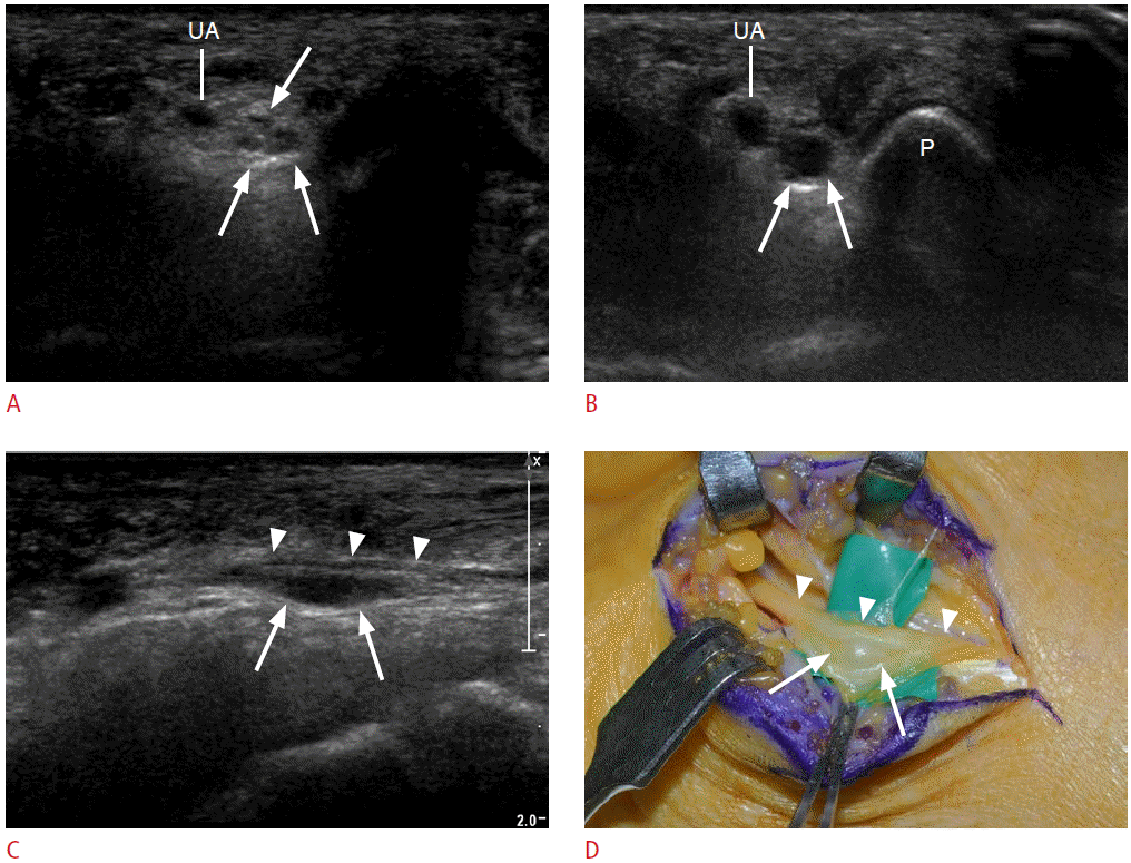
Fig. 14.
The schematic representation and transverse sonograms of the normal radial nerve at the level of the spiral groove in the posterior arm.
A. Dashed arrows inserted in the schematic indicate the corresponding levels of the sonograms. B-D. Consecutive sonograms from the proximal to distal posterior arm. Color Doppler ultrasonography (B) can identify the radial nerve (arrow) at the level of the proximal posterior arm. Note the course of the radial nerve from the proximal medial (arrow in B) to the distal lateral (arrow in D) of the posterior arm. Note the close proximity to the posterior humeral shaft.
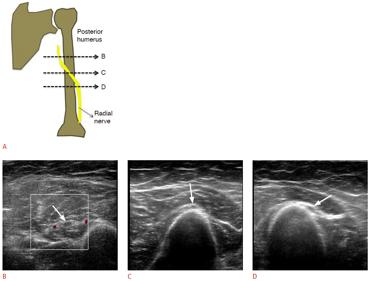
Fig. 15.
The schematic representation of the radial tunnel.
The radial nerve bifurcates into the superficial branch and the deep motor branch at the level of the capitellum of the humerus. The radial tunnel is where the deep motor branch of the radial nerve lies; from the bifurcation point of the radial nerve (or the radiocapitellar joint) to the upper edge of the supinator muscle (arcade of Frohse). ECRB, extensor carpi radialis brevis muscle; ECRL, extensor carpi radialis longus muscle.
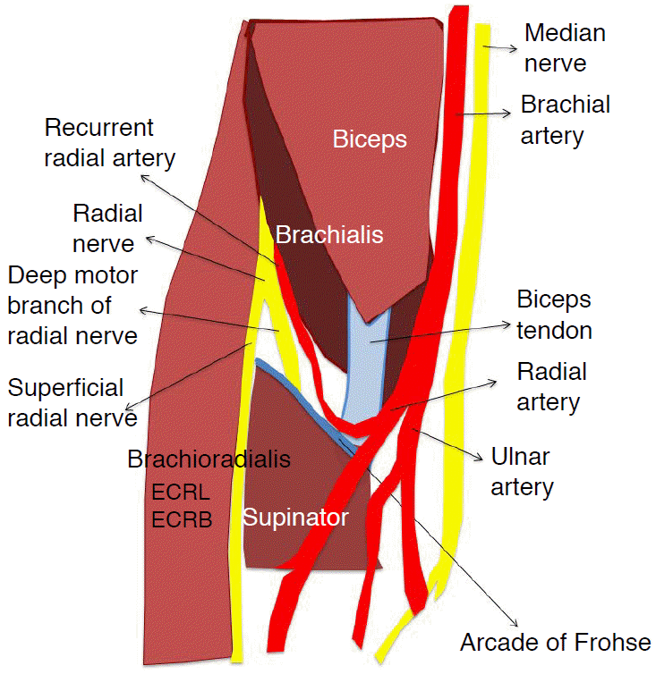
Fig. 16.
Sonograms of the normal radial nerve at the level of the radial tunnel.
A. The transverse color Doppler sonogram at the level of the elbow joint shows two branches of the radial nerve (arrows) in the muscle fascial plane between brachioradialis (BR) and brachialis (B) muscles. C, capitellum of the humerus. B, C. Transverse sonograms at the level of the distal radial tunnel (B) and the supinator muscle (C) show the deep motor branch of the radial nerve (arrow) entering the supinator muscle (S), whereas the superficial radial nerve (arrowhead) is running along the deep portion of the BR. D. More distally, the transverse sonogram of the lateral forearm shows the deep motor branch of the radial nerve exiting the supinator muscle (S) as the posterior interosseous nerve (arrow). E. The longitudinal sonogram of the radial tunnel shows the deep motor branch of the radial nerve (arrowheads) entering the supinator muscle (S). Arrows indicate the level of the upper edge of the supinator muscle (S); the arcade of Frohse.
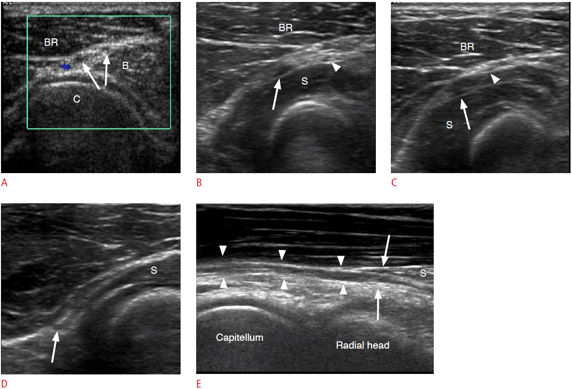
Fig. 17.
The schematic representation and transverse sonograms of the relationship between the superficial radial nerve and the extensor tendons.
A. Dashed arrows inserted in the schematic indicate the corresponding levels of the sonograms. 1st MC, 1st metacarpal bone; APL, abductor pollicis longus tendon; Distal R, distal radius; ECRB, extensor carpi radialis brevis tendon; ECRL, extensor carpi radialis longus tendon; EPB, extensor pollicis brevis tendon; EPL, extensor pollicis longus tendon. B. The transverse sonogram at the level of the distal radius shows the superficial radial nerve (arrow) and cephalic vein (arrowhead), overlying the first extensor compartment tendons (APL and EPB). C. More distally, the superficial radial nerve (arrow) crossed over the first extensor compartment tendons (APL and EPB) and is seen between extensor compartment I (APL and EPB) and II (ECRL and ECRB). The cephalic vein is collapsed due to compression by the transducer (arrowhead).
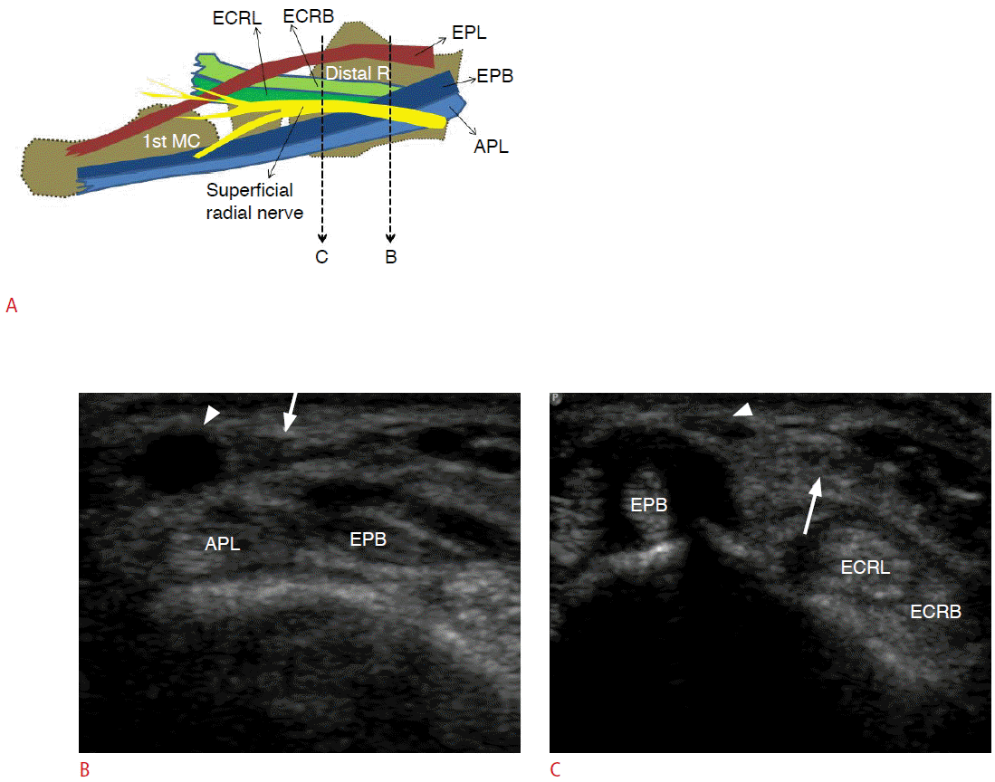
Fig. 18.
A 61-year-old female patient with radial tunnel syndrome.
The patient presented with lateral elbow pain without motor weakness. A. The transverse sonogram shows a ganglionic cyst in the radial tunnel compressing the deep motor branch (white arrow) and the superficial branch (black arrow) of the radial nerve. Note the adjacent recurrent radial artery (arrowhead). B. The longitudinal sonogram reveals the displaced deep motor branch of the radial nerve (arrows) by the ganglionic cyst within the radial tunnel.

- REFERENCES
- REFERENCES
References
1. Goedee HS, Brekelmans GJ, van Asseldonk JT, Beekman R, Mess WH, Visser LH. High resolution sonography in the evaluation of the peripheral nervous system in polyneuropathy: a review of the literature. Eur J Neurol 2013;20:1342–1351.
[Article] [PubMed]2. Cartwright MS, Walker FO. Neuromuscular ultrasound in common entrapment neuropathies. Muscle Nerve 2013;48:696–704.
[Article] [PubMed]3. Miller TT, Reinus WR. Nerve entrapment syndromes of the elbow, forearm, and wrist. AJR Am J Roentgenol 2010;195:585–594.
[Article] [PubMed]4. Andreisek G, Crook DW, Burg D, Marincek B, Weishaupt D. Peripheral neuropathies of the median, radial, and ulnar nerves: MR imaging features. Radiographics 2006;26:1267–1287.
[Article] [PubMed]5. Jacobson JA, Fessell DP, Lobo Lda G, Yang LJ. Entrapment neuropathies I: upper limb (carpal tunnel excluded). Semin Musculoskelet Radiol 2010;14:473–486.
[Article] [PubMed]6. Kim SJ, Hong SH, Jun WS, Choi JY, Myung JS, Jacobson JA, et al. MR imaging mapping of skeletal muscle denervation in entrapment and compressive neuropathies. Radiographics 2011;31:319–332.
[Article] [PubMed]7. Ulrich D, Piatkowski A, Pallua N. Anterior interosseous nerve syndrome: retrospective analysis of 14 patients. Arch Orthop Trauma Surg 2011;131:1561–1565.
[Article] [PubMed] [PMC]8. Hill NA, Howard FM, Huffer BR. The incomplete anterior interosseous nerve syndrome. J Hand Surg Am 1985;10:4–16.
[Article] [PubMed]9. Klauser AS, Faschingbauer R, Bauer T, Wick MC, Gabl M, Arora R, et al. Entrapment neuropathies II: carpal tunnel syndrome. Semin Musculoskelet Radiol 2010;14:487–500.
[Article] [PubMed]10. Beekman R, Visser LH. Sonography in the diagnosis of carpal tunnel syndrome: a critical review of the literature. Muscle Nerve 2003;27:26–33.
[Article] [PubMed]11. Tajika T, Kobayashi T, Yamamoto A, Kaneko T, Takagishi K. Diagnostic utility of sonography and correlation between sonographic and clinical findings in patients with carpal tunnel syndrome. J Ultrasound Med 2013;32:1987–1993.
[Article] [PubMed]12. Mallouhi A, Pulzl P, Trieb T, Piza H, Bodner G. Predictors of carpal tunnel syndrome: accuracy of gray-scale and color Doppler sonography. AJR Am J Roentgenol 2006;186:1240–1245.
[Article] [PubMed]13. Wong SM, Griffith JF, Hui AC, Lo SK, Fu M, Wong KS. Carpal tunnel syndrome: diagnostic usefulness of sonography. Radiology 2004;232:93–99.
[Article] [PubMed]14. Sernik RA, Abicalaf CA, Pimentel BF, Braga-Baiak A, Braga L, Cerri GG. Ultrasound features of carpal tunnel syndrome: a prospective case-control study. Skeletal Radiol 2008;37:49–53.
[Article] [PubMed]15. Kim HS, Joo SH, Cho HK, Kim YW. Comparison of proximal and distal cross-sectional areas of the median nerve, carpal tunnel, and nerve/tunnel index in subjects with carpal tunnel syndrome. Arch Phys Med Rehabil 2013;94:2151–2156.
[Article] [PubMed]16. Bianchi S, Martinoli C. Wrist. Bianchi S, Martinoli C. Ultrasound of the musculoskeletal system. Berlin: Springer, 2007:425–494.17. Buchberger W, Judmaier W, Birbamer G, Lener M, Schmidauer C. Carpal tunnel syndrome: diagnosis with high-resolution sonography. AJR Am J Roentgenol 1992;159:793–798.
[Article] [PubMed]18. Duncan I, Sullivan P, Lomas F. Sonography in the diagnosis of carpal tunnel syndrome. AJR Am J Roentgenol 1999;173:681–684.
[Article] [PubMed]19. Buchberger W, Schon G, Strasser K, Jungwirth W. High-resolution ultrasonography of the carpal tunnel. J Ultrasound Med 1991;10:531–537.
[Article] [PubMed]20. Ozturk E, Sonmez G, Colak A, Sildiroglu HO, Mutlu H, Senol MG, et al. Sonographic appearances of the normal ulnar nerve in the cubital tunnel. J Clin Ultrasound 2008;36:325–329.
[Article] [PubMed]21. Apfelberg DB, Larson SJ. Dynamic anatomy of the ulnar nerve at the elbow. Plast Reconstr Surg 1973;51:79–81.
[Article]22. Volpe A, Rossato G, Bottanelli M, Marchetta A, Caramaschi P, Bambara LM, et al. Ultrasound evaluation of ulnar neuropathy at the elbow: correlation with electrophysiological studies. Rheumatology (Oxford) 2009;48:1098–1101.
[Article] [PubMed]23. Wiesler ER, Chloros GD, Cartwright MS, Shin HW, Walker FO. Ultrasound in the diagnosis of ulnar neuropathy at the cubital tunnel. J Hand Surg Am 2006;31:1088–1093.
[Article] [PubMed]24. Thoirs K, Williams MA, Phillips M. Ultrasonographic measurements of the ulnar nerve at the elbow: role of confounders. J Ultrasound Med 2008;27:737–743.
[Article] [PubMed]25. Yoon JS, Walker FO, Cartwright MS. Ultrasonographic swelling ratio in the diagnosis of ulnar neuropathy at the elbow. Muscle Nerve 2008;38:1231–1235.
[Article] [PubMed]27. Gross MS, Gelberman RH. The anatomy of the distal ulnar tunnel. Clin Orthop Relat Res 1985;(196):238–247.
[Article]28. Pierre-Jerome C, Moncayo V, Terk MR. The Guyon's canal in perspective: 3-T MRI assessment of the normal anatomy, the anatomical variations and the Guyon's canal syndrome. Surg Radiol Anat 2011;33:897–903.
[Article] [PubMed]29. Zeiss J, Jakab E, Khimji T, Imbriglia J. The ulnar tunnel at the wrist (Guyon's canal): normal MR anatomy and variants. AJR Am J Roentgenol 1992;158:1081–1085.
[Article] [PubMed]30. Thomas SJ, Yakin DE, Parry BR, Lubahn JD. The anatomical relationship between the posterior interosseous nerve and the supinator muscle. J Hand Surg Am 2000;25:936–941.
[Article] [PubMed]31. De Maeseneer M, Marcelis S, Jager T, Girard C, Gest T, Jamadar D. Spectrum of normal and pathologic findings in the region of the first extensor compartment of the wrist: sonographic findings and correlations with dissections. J Ultrasound Med 2009;28:779–786.
[Article] [PubMed]32. Tagliafico A, Cadoni A, Fisci E, Gennaro S, Molfetta L, Perez MM, et al. Nerves of the hand beyond the carpal tunnel. Semin Musculoskelet Radiol 2012;16:129–136.
[Article] [PubMed]33. Djurdjevic T, Loizides A, Loscher W, Gruber H, Plaikner M, Peer S. High resolution ultrasound in posterior interosseous nerve syndrome. Muscle Nerve 2014;49:35–39.
[Article] [PubMed]34. Ong C, Nallamshetty HS, Nazarian LN, Rekant MS, Mandel S. Sonographic diagnosis of posterior interosseous nerve entrapment syndrome. Radiol Case Rep 2007;2:67.
[Article]35. Umehara F, Yoshino S, Arimura Y, Fukuoka T, Arimura K, Osame M. Posterior interosseous nerve syndrome with hourglass-like fascicular constriction of the nerve. J Neurol Sci 2003;215:111–113.
[Article] [PubMed]36. Chien AJ, Jamadar DA, Jacobson JA, Hayes CW, Louis DS. Sonography and MR imaging of posterior interosseous nerve syndrome with surgical correlation. AJR Am J Roentgenol 2003;181:219–221.
[Article] [PubMed]
