

 Print
Print facebook
 twitter
 Linkedin
 google+

Introduction
Imaging-guided core needle biopsy (CNB) is becoming the standard of diagnosis for breast disease as a reliable alternative to open excision biopsy [1-5]. Percutaneous image-guided CNB is performed under either stereotactic or ultrasonographic guidance. Percutaneous ultrasound (US)-guided breast biopsy has several advantages over stereotactic or surgical biopsy, and many studies have reported CNB to be effective and safe. This procedure is safe; faster; less expensive; is performed in real time, allowing accurate assessments; and does not involve ionizing radiation [4,6,7].
However, the possibility of false-negative results is unavoidable; false-negatives can result from core needle sampling errors, failure to recognize imaging-histology discordances, and inappropriate follow-up periods for benign biopsies [8,9]. A delayed diagnosis of breast cancer is an outcome that every clinician strives to avoid. In previous studies of US-guided 14-gauge CNBs with at least 2 years of follow-up, the false-negative rates has ranged from 0.1% to 2.5% [5,10-12]. So far, some studies have been published on US-guided CNB for breast lesions with long-term follow-up periods [5,6,13,14], but most previous reports of US-guided CNB in the breast were based on a small number of biopsies. Furthermore, few studies have reported large series of US-guided CNB with long-term follow-up; therefore, this study was undertaken, based on a relatively large series of US-guided 14-gauge CNB procedures performed for breast lesions with long-term follow-up periods.
The purpose of this study was to assess the outcomes of US-guided CNB for breast lesions that had at least a 2-year follow-up for benign lesions in a large series to determine the false-negative rate and to evaluate the diagnostic performance of CNB.
Materials and Methods
Study Population
This study was conducted with Institutional Review Board approval, and informed consent was waived because of the retrospective nature of this study.
From July 2005 to December 2012, 13,254 consecutive US-guided 14-gauge CNBs for breast lesions were performed at our institution. We retrospectively reviewed all the biopsy results and excluded 4,186 biopsies for which a non-malignant biopsy result was not confirmed by surgical excision or US-guided vacuum-assisted biopsy, or at least 2 years of follow-up data were not available.
A total of 9,068 breast masses in 7,039 women (age range, 11 to 92 years; mean, 46 years) were included in this study. Of the 7,039 patients, 1,253 had biopsies of two separate lesions, 234 had biopsies of three separate lesions, 74 had biopsies of four separate lesions, 11 had biopsies of five separate lesions, four had biopsies of six separate lesions, one had biopsies of seven separate lesions, one had biopsies of eight separate lesions, and one had biopsies of 10 separate lesions.
Biopsy Procedure
US-guided 14-gauge CNB was performed using a free-hand technique and a high-resolution US unit with 7.5- or 12-MHz linear array transducers (HDI 5000 or IU22, Philips-Advanced Technology Laboratories, Bothell, WA, USA; or Logiq 9, GE Healthcare, Milwaukee, WI, USA). Each procedure was performed in an outpatient setting under local anesthesia with the patient in the supine position. A 14-gauge automated core biopsy needle with a spring-loaded biopsy gun (Promac 2.2L, Manan Medical Products, Northbrook, IL, USA) or a 14-gauge dual-action semiautomatic core biopsy needle with a 22-mm throw (Stericut cut with coaxial, TSK Laboratory, Tochigi, Japan) was used. All biopsies were performed by one of 19 radiologists in fellowship training (n=15) or with extensive clinical experience (n=4) who were specialists in breast imaging and biopsies. According to our standard protocol, four or five core samples per lesion were routinely obtained.
Post-biopsy Procedure
For each lesion that underwent a CNB, a radiologist reviewed the pathology report in conjunction with the images obtained before, during, and after the biopsy procedure, and based on the results of the review, an addendum was attached to the biopsy report recommending specific management strategies for the patients and the referring physicians [15,16]. The imaging and histological findings were considered to be concordant when the histological findings provided a sufficient explanation for the imaging findings and discordant when they did not [15]. For malignant lesions (e.g., invasive carcinoma, ductal carcinoma in situ [DCIS], and metastases) identified through 14-gauge CNB, immediate definitive surgery or chemotherapy was recommended, in accordance with what was deemed clinically appropriate. For high-risk lesions (e.g., atypia including atypical ductal hyperplasia [ADH], lobular neoplasia, radial sclerosing lesions, and possible phyllodes tumors) and benign lesions (neither malignant nor high-risk) with imaging-histology discordance (i.e., a lesion that was thought to be malignant based on imaging but was demonstrated to be benign by histological findings) resulted in recommendations for surgical excision [15,17].
US follow-up at 6 months after biopsy and then annually for at least 2 years was recommended in patients with concordant benign lesions (i.e., a lesion that was suspected of being benign based on imaging and also proven to be benign by the histological findings). For some concordant benign lesions, rebiopsies were carried out by surgical excision or US-guided vacuum-assisted biopsy at the request by the patient or referring physician or because of suspicious physical findings (i.e., a palpable mass or nipple discharge) or lesion progression at US follow-up. The choice of surgical excision or US-guided vacuum-assisted biopsy at rebiopsy depended on the patient’s or referring physician’s preferences.
Data Analysis
The pathologic findings of US-guided 14-gauge CNBs and rebiopsies, as well as the follow-up results, were obtained from patients’ medical records. After reviewing the results, pathologic comparisons between US-guided 14-gauge CNB results and the standard diagnostic reference were made. The standard diagnostic reference included the results of surgical excision, vacuum-assisted biopsy, or at least 2 years of long-term imaging follow-up. The false-negative rate was calculated as the proportion of benign results from US-guided 14-gauge CNBs among all breast cancers [5,10]. The underestimation rate for high-risk lesions was calculated as the proportion of lesions diagnosed as high-risk by CNB that were finally proven to be DCIS or invasive cancer after surgical excision. The underestimation rate for DCIS was likewise defined as the proportion of lesions diagnosed as DCIS by CNB that were finally proven to be invasive cancer after surgical excision [5]. For the false-negative results of US-guided 14-gauge CNB, the time interval between the CNB and rebiopsy, as well as the reasons for rebiopsy, were reviewed.
Results
The lesions ranged in size from 2 to 130 mm (mean, 14.0 mm) as measured by US. The distribution of size and Breast Imaging-Reporting and Data System (BI-RADS) categories are summarized in Table 1. Of 9,068 breast masses, 4,408 lesions (48.6%) were less than 10 mm, and the most common indication for biopsy was the presence of a BI-RADS category 4a lesion (54.0%, 4,900 of 9,068). Three CNBs were BI-RADS category 1; among these, two cases were regarded as normal breast tissue on US, but the patient requested a biopsy. The remaining such case was a patient who had undergone a mastectomy and showed normal breast tissue on US, but a positron emission tomography scan revealed increased fluorodeoxyglucose uptake. All three cases were identified as benign on pathologic reports after the biopsies.
Pathologic Results and Underestimation Rate of CNB
Among the 9,068 breast masses, the pathologic results of US-guided 14-gauge CNB were benign in 64.2% (5,821 lesions), high-risk in 3.5% (322 lesions), and malignant in 32.3% (330 DCIS lesions and 2,595 cases of invasive cancer) (Tables 2, 3). A total of 4,782 cases were confirmed by surgical excision or US-guided vacuum-assisted biopsy; in these cases, breast cancer was found in 3.2% (63 of 1,990) of benign CNBs, 26.3% (79 of 300) of high-risk CNBs, and 100% (2,492 of 2,492) in malignant CNBs at the final diagnosis. The malignancy rates were 0% (0 of 3) for lesions in BI-RADS category 1, 2.8% (1 of 35) in category 2, 2.2% (28 of 1,248) in category 3, 9.4% (463 of 4,900) in category 4a, 47.7% (267 of 560) in category 4b, 83.9% (640 of 762) in category 4c, and 97.8% (1,526 of 1,560) in category 5. The underestimation rate was 33.6% (111 of 330) for DCIS and 24.5% (79 of 322) for high-risk lesions at surgical excision (Fig. 1).
False-Negative Results
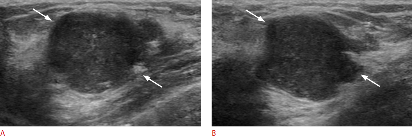
Of the 5,821 benign CNB results, 63 lesions were confirmed to be malignant after surgical excision (1.1%, 63 of 5,821). After postbiopsy review, 5,586 benign CNBs showed imaging-pathology concordance, which included 29 lesions that were ultimately diagnosed as malignant (0.5%, 29 of 5,586), while 235 benign CNBs showed imaging-pathology discordance (Fig. 2), including 34 lesions determined to be malignant after the subsequent surgical excision (14.4%, 34 of 235). A total of 3,067 cases were malignant at the final diagnosis, and the false-negative rate was 2.0% (63 of 3,067) with a sensitivity of 95.4% (2,927 of 3,067). Of the false-negative results, papillary lesions represented the highest percentage (36.5%, 23 of 63), followed by fibrocystic changes (20.6%, 13 of 63) and fibrosis (9.5%, 6 of 63) (Table 2). Most false-negative diagnoses (84.1%, 53 of 63) underwent immediate rebiopsy (interval range, 3 to 79 days; mean, 31.1 days) because the radiologist noted the discordance between the imaging findings and CNB and recommended surgical excision or a vacuum-assisted biopsy.
Analysis of Delayed False-Negative Diagnoses
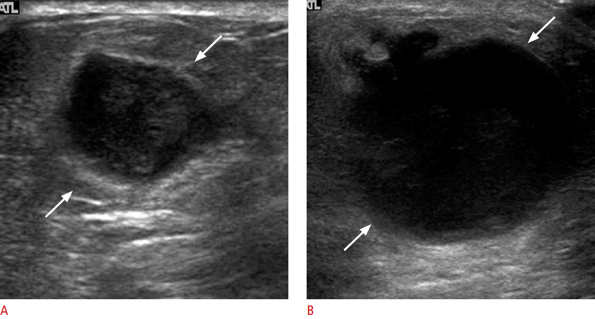
Ten malignancies (15.9%, 10 of 63) had delayed diagnoses, and the mean time interval between the initial CNB and excision was 11.6 months (range, 5 to 17 months) (Table 4). For five benign lesions, although radiologists performed imaging-pathology correlations and recommended surgical excision immediately after the initial CNB, the patients refused excision, resulting in delayed diagnoses of malignancy. All 10 delayed false-negative diagnoses showed progression in follow-up US imaging (Fig. 3). Of the five cases in which excision was refused, one lesion (Table 4, patient 1) was categorized as BI-RADS category 4a and the result of the initial US-guided CNB was fibrocystic change, which was considered to be concordant. However, a radiologist recommended surgical excision because of its large size (30 mm); the lesion increased in size from 30 to 43 mm over 17 months, and was confirmed as a malignant phyllodes tumor following surgical excision. Another patient (Table 4, patient 8) had a history of breast surgery due to intraductal papilloma 13 years ago, and a new mass was categorized as BI-RADS category 4a. The result of CNB was focal fibrosis, but a radiologist concluded that the lesion had high possibility of being intraductal papilloma and recommended surgical excision. The lesion markedly increased in size from 24 to 50 mm over 17 months, and was confirmed as invasive lobular carcinoma following surgical excision. Both of these patients refused excision after the initial CNB, causing delayed diagnoses of malignancy.
Discussion
In this study, we analyzed a large population in which US-guided CNBs of breast lesions were performed from July 2005 to December 2012, building upon the previous study conducted from February 2000 through June 2005 at our institution [5]. The inclusion criteria were the same in both studies, and we included a total of 9,068 breast masses in this study, in contrast to 2,420 masses in the previous study. The mean lesion size was smaller in this study (mean, 14.0 mm; range, 2 to 130 mm) than in the previous study (mean, 18.7 mm; range, 2 to 180 mm), but we showed a lower false negative rate of 2.0% (63 of 3,067) than the false-negative rate of 2.4% (31 of 1,312) observed in the previous study. Overall, the results of our study showed that US-guided 14-gauge CNB for breast lesions exhibited better performance, based on the analysis of a larger population with smaller lesions in contrast to the sample of the preceding study from the same institution.
False-negative diagnoses are unavoidable for reasons such as sampling errors, failure to act upon imaging-histology discordances, or the absence of imaging follow-up after a benign biopsy [9]. However, most false-negative diagnoses can be immediately recognized through careful post-biopsy review, as was exemplified by our data. Most of our false-negative results (84.1%, 53 of 63) underwent immediate rebiopsies, avoiding delayed diagnoses of cancer, because imaging-pathology discordances were promptly detected by radiologists. Previous studies have shown similar results [5,6,13,18]. Several studies have suggested that physicians should perform rebiopsies or surgical excision for discordant CNBs because of the high prevalence of carcinoma in imaging-pathology discordant lesions [15,19,20]. Our data likewise showed a much higher malignant rate of 14.4% in discordant benign CNBs, which is within the 6.8%-24.4% range reported in previous studies [15,19,21], compared with a malignancy rate of 0.5% in concordant benign CNBs. These results accentuate the importance of imaging-pathology correlations after biopsy and the significance of discordant lesions.
Of the false-negative diagnoses, papillary lesions comprised the largest subgroup (36.5%, 23 of 63) in terms of the histological classification, and 3.1% (23 of 728) of the papillary lesions that underwent CNB were upgraded to malignancy; this is comparable with previous studies, in which 2.3%-14% of papillary lesions were upgraded to DCIS or invasive cancer after surgical excision [22-26]. To decide whether a papillary lesion is benign or malignant on the basis of CNB is challenging because of the heterogeneity of papillomas and targeting error [27,28]. It remains controversial whether all benign papillary lesions on CNB should undergo surgical excision to avoid false-negative diagnoses. Our data showed an upgrade rate to carcinoma of only 2.3% (16 of 686) in concordant papillomas, in contrast to an upgrade rate of 16.6% (7 of 42) in discordant papillomas; this supports the suggestions of recent reports that observation is sufficient, rather than surgical excision, for papillomas identified as benign by CNB if the imaging-pathology findings are concordant [26,29].
Ten malignancies had delayed diagnoses without immediate rebiopsies in the false-negative results. All these lesions showed progression in follow-up US. After review of the patients’ medical records, we found that patients refused excision for five false-negative lesions, although radiologists performed imaging-pathology correlations for these lesions and recommended surgical excision immediately after the initial CNB. The other five delayed false-negative diagnoses were identified as malignancies on follow-up sonography within 1 year after the initial CNB. Our results suggest that it is important to consider not only appropriate follow-up, but patient compliance after the biopsy. In previous studies, compliance rates of only 54% (49 of 90 lesions) [30] and 50.9% (84 of 165 lesions) [31] were reported for imaging surveillance recommendations. Therefore, poor patient compliance and inappropriate follow-up may cause further delays in the diagnosis and treatment of breast cancer, even if imaging-pathology discordances are recognized immediately.
One of the limitations of CNB is the histologic underestimation of breast malignancy, which means that lesions found to be high-risk or DCIS by a percutaneous breast biopsy are upgraded to DCIS or invasive cancer after surgical excision. In previous studies, the underestimation rates ranged from 6.25% to 65% for ADH and from 16% to 66% for DCIS using CNB or vacuum-assisted biopsy [13,32-35]. Our underestimation rates were 24.5% for high-risk lesions (79 of 322) and 33.6% for DCIS (111 of 330), comparable with previous reports.
Our study has a few limitations. First, there was no retrospective pathologic review of the core needle samples for the false-negative results. Second, the radiologists who performed the biopsies were heterogeneous, and included radiologists undergoing fellowship training (n=15). However, our institution is a tertiary hospital and it is natural for less experienced trainees to perform biopsies. Furthermore, our results showed reliable sensitivity (95.4%) and a low false-negative rate (2.0%).
In conclusion, US-guided 14-gauge CNB is accurate and provides optimal diagnostic information for breast lesions. Imaging-histology correlations and appropriate imaging follow-up should be performed to identify possible false-negative results and to avoid delayed diagnoses.

 Download Citation
 PDF Links
 PubReader
 ePub Link
 Full text via DOI
 Full text via PMC


