

 Print
Print facebook
 twitter
 Linkedin
 google+

Introduction
Pediatric foot pain can stem from various etiologies. An accurate diagnosis requires a comprehensive understanding of pediatric foot anatomy, growth patterns, and biomechanics, along with a thorough medical history and physical examination in clinical practice to identify the underlying cause. Musculoskeletal ultrasonography (US) offers real-time visualization of both bone surfaces and the surrounding soft tissue from multiple angles, providing superior detail for determining the etiology of foot pain and tracking disease progression compared to alternative imaging modalities. Given the potential for misleading clinical diagnoses based solely on symptoms and physical examination, US has emerged as a valuable problem-solving tool to make an accurate diagnosis. Furthermore, understanding the anatomical location of the pain can assist radiologists in conducting US examinations efficiently and effectively, thereby narrowing down the differential diagnosis (Fig. 1).
This review presents common foot disorders in children and adolescents, summarizing their US manifestations. By harnessing the convenience, speed, few contraindications, real-time and high resolution of high-frequency US examinations, it is hoped that the information presented herein will provide powerful support for the diagnosis of these conditions.
Kohler’s Disease
Epidemiology
Kohler’s disease, also known as osteochondrosis of the tarsal navicular bone, was originally described by Kohler in 1908. Kohler’s disease is a rare condition that mostly affects children younger than 10 years [1]. The overall incidence is higher in males than in females, and it is bilateral in up to 25% of cases [2,3].
Pathophysiology
Most cases of Kohler’s disease are idiopathic [4]. The etiology may be attributed to the avascular necrosis of the navicular bone due to a mechanical insult caused by external forces. The navicular bone bears weight, is the latest-ossifying bone among all the tarsal bones, and serves as the apex of the medial longitudinal arch. The ossification center of the navicular bone may suffer compression force trauma as a result of delayed ossification or mechanical fatigue, eventually leading to avascular navicular necrosis when the intraosseous blood flow is compromised.
Clinical Findings
There is often no specific eliciting trigger in Kohler’s disease. Pain in the area of the navicular bone that worsens with weight bearing is a common presenting complaint of the disease. The patients may be found to walk with an antalgic gait that favors the lateral border of the foot [5,6] and might present with a limp. On physical examination, focal midfoot tenderness and swelling may be seen, and the pain increases in intensity with posterior tibial tendon traction.
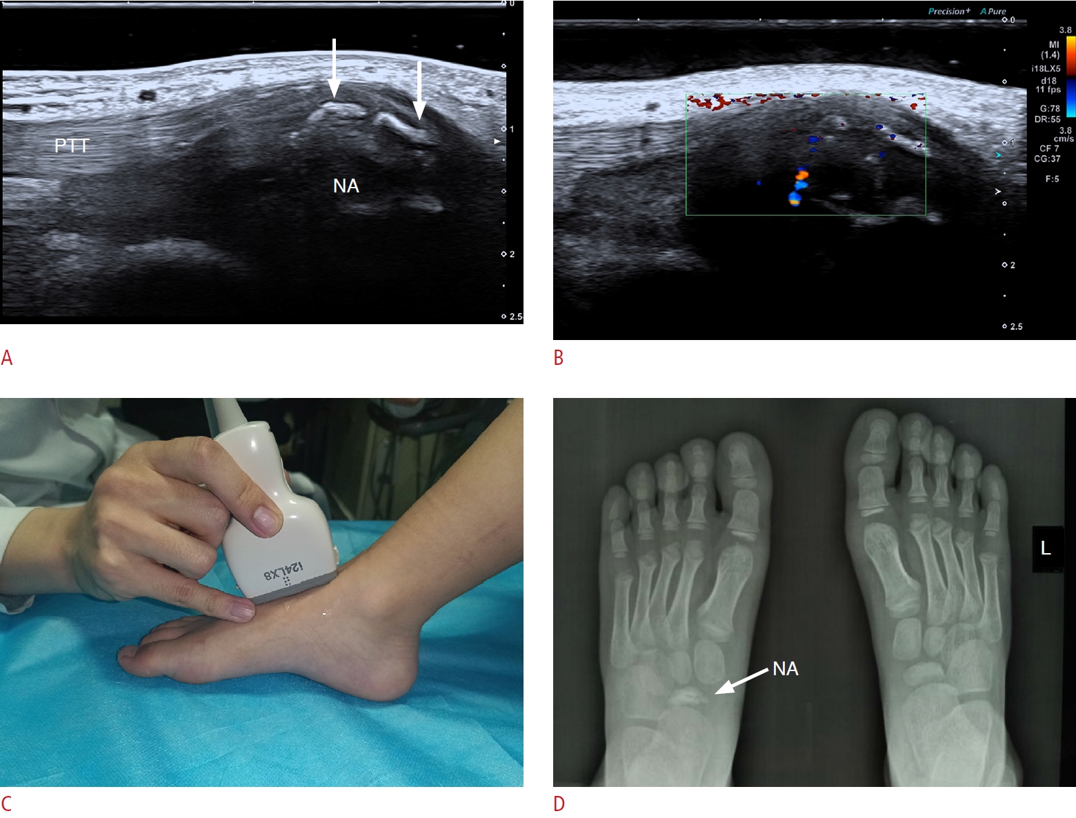
Ultrasound Findings
To evaluate the navicular bone, it is recommended for the patient to sit or lie in a supine position with the foot flat on the bed.
US shows that the surface of the navicular ossification center is unsmooth, bumpy, fragmented and even deformed. Posterior tibialis tendonitis and/or tenosynovitis may accompany Kohler’s disease (Fig. 2). Comparing the affected and unaffected sides and eliciting symptoms by applying pressure to the affected area using the US transducer can enhance diagnostic certainty.
Chan and Young [6] reported that the diagnosis of Kohler’s disease primarily relies on X-ray or magnetic resonance imaging (MRI). The most common pattern involves fragmentation and flattening of the navicular bone, which is often described as "waferlike," with patchy areas of sclerosis and loss of the normal bony trabecular architecture. The case presented in Fig. 2 provides evidence that US, similar to X-ray imaging, can display the abnormal appearance of the ossification center within the navicular bone. However, there is a lack of adequate US findings previously documented for Kohler's disease in the literature.
Treatment
Kohler’s disease is a self-limiting condition without long-term complications. The mainstay of management is non-operative, involving a cast for immobilization for 4 to 6 weeks to help mitigate symptoms [1]. Karr [7] demonstrated that external fixation with navicular diastasis is an effective, straightforward management option that allows osseous regrowth and bone healing.
Sever’s Disease
Epidemiology
Sever’s disease, or calcaneal apophysitis, is the most common cause of heel pain in pediatric patients, and its incidence ranges from 2% to 16% in children [8,9]. It occurs in the area where the Achilles tendon attaches to the calcaneus. Sever's disease is an overuse syndrome that is frequently observed in athletic pediatric patients between the ages of 8 and 12 years [10,11]. Inadequate footwear, early specialization in sports, and contracture of the Achilles tendon may contribute to the development of the disease [2,9].
Pathophysiology
The calcaneus growth plate appears at the age of 8 and does not close until at least 14 years of age. The most significant etiological factor for injuries leading to Sever’s disease is mechanical overuse and microtrauma due to repetitive impact pressure and tractive shear stresses to this open growth plate of the calcaneus. A prospective study evaluating 14 patients with Sever’s disease found that their post-treatment MRI findings demonstrated resolution of the metaphyseal manifestations, suggesting that this disease may be related to increased metaphyseal stress as a result of biomechanical changes [9].
Clinical Findings
Children often complain of pain over the posterior heel at the insertion point of the Achilles tendon, and physical activities can worsen the pain. Patients often walk on their toes to avoid putting weight on heels. A physical examination demonstrates tenderness with palpation and compression at the medial and lateral aspects of the heel [2], and there is typically no heel swelling. A clinical diagnosis can be made by performing a "squeeze test" of the heel [12,13]. In addition, Sever's disease complicated by calcaneal osteomyelitis is a serious condition that can lead to severe complications [14,15].
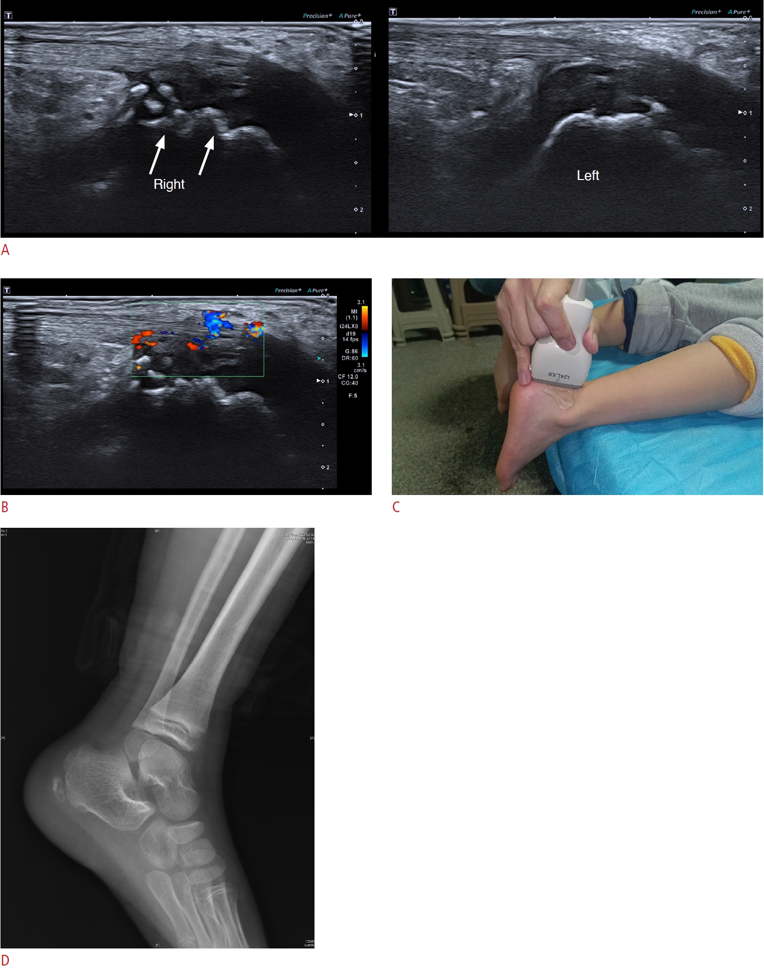
Ultrasound Findings
A US examination is usually performed with the patient lying on the bed in a prone position with the patient’s feet hanging over the edge of the bed [16].
US shows fragmentation of the secondary nucleus of the calcaneus, the surface of which can be rough, serrated, fissured [17], and deformed [18]. Achilles tendinitis may occur in conjunction with Sever’s disease, with US findings of tendon thickening, hypo-echogenicity and indistinct fiber structures at the area of insertion into the calcaneus apophysis. Dot-shaped and short rod-shaped blood flow signals in tendon lesions indicate active inflammation [19]. The presence of retrocalcaneal bursitis is also considered characteristic [17]. Bursitis presents most often as an enlarged bursa filled with anechoic exudates on US (Fig. 3).
Volpon and de Carvalho Filho [20] conducted a study involving 392 children and found that the fragmentation of the calcaneal apophysis represented a significant radiographic finding, a conclusion that aligns with the US observations reported by Hosgoren et al. [17]. MRI can also diagnose calcaneal apophysitis clearly.
Treatment
Sever’s disease is a self-limiting disorder that resolves when the apophyseal centers fuse [21]. Treating Sever’s disease consists of a combination of multiple treatment modalities. Activity restriction, icing, heel cord stretching, immobilization, nonsteroidal anti-inflammatory drugs (NSAIDs), heel cups [22,23], and an X-brace [24] can be utilized to relieve pain [8,25].
Iselin’s Disease
Epidemiology
Iselin’s disease was first described by Dr. Hans Iselin in 1912. It refers to a painful traction apophysitis affecting the tuberosity of the base of the fifth metatarsal. Iselin’s disease most commonly occurs in young athletic adolescents [2]. Iselin’s disease is uncommon but not rare in clinical practice; however, the currently available published data are limited [26]. Apophysitis refers to an injury within the peroneus brevis tendon insertion site and appears approximately at age 10 to 11 years in females and 12 to 14 years in males, consistent with the appearance of the proximal apophysis of the fifth metatarsal tuberosity.
Pathophysiology
The condition can be caused by repetitive microtrauma through the tractive force of the peroneus brevis tendon acting on the base of the fifth metatarsal, resulting in inflammation and even avulsion of the apophysis.
Clinical Findings
The most common symptom is pain in the lateral part of the midfoot [5] or around the base of the fifth metatarsal, with symptoms aggravated by activities or wearing shoes. The physical examination findings include swelling and tenderness over the fifth metatarsal area (relative to the contralateral foot) on palpation and resisted inversion. Additionally, symptoms may be elicited with eversion and plantar flexion. The pain usually disappears when apophysis fusion occurs.
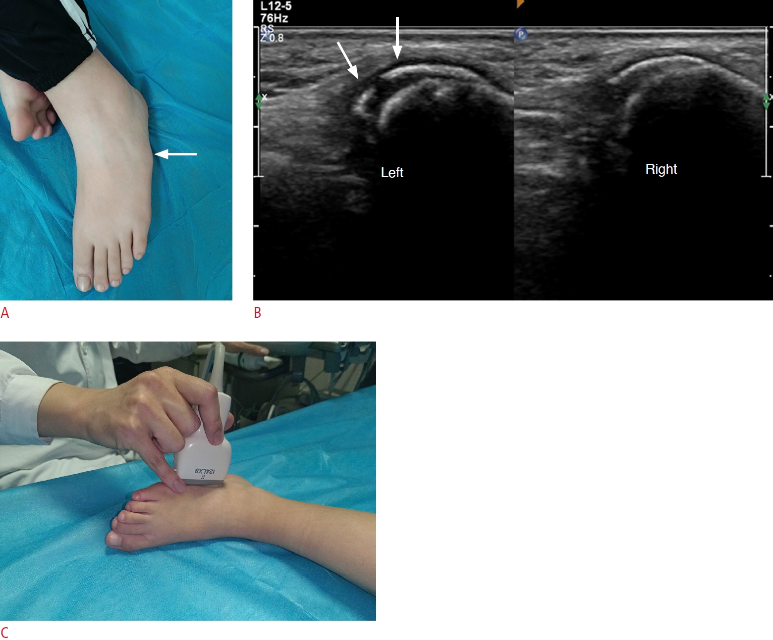
Ultrasound Findings
The optimal patient position involves either sitting or lying supine with the foot flat on the bed and inverted in the reverse frog-leg position for evaluating the base of the fifth metatarsal.
US shows a thickened epiphysis and irregular or fragmented ossification center of the base of the fifth metatarsal. The peroneus brevis tendon involved in Iselin’s disease is thickened and hypoechoic, with increased blood flow (Fig. 4).
Limited descriptions of the US features of this condition are available in the literature. Forrester et al. [26] summarized the radiological manifestations of Iselin's disease, including enlargement and widening of the apophysis, as well as fragmentation of the ossification center. Kishan et al. [27] proposed that MRI could potentially reveal bone marrow edema of the fifth metatarsal.
Treatment
Treatment strategies for Iselin’s disease involve ice packs, restriction of sports activities, and analgesics. Short leg casts or walking boot immobilization and physical therapy may be required while severe symptoms persist [28]. Symptomatic nonunion may require surgical excision of the disrupted bone [29].
Talocalcaneal Coalition
Epidemiology
Tarsal coalition was first described in 1769. The disorder is one of the most common causes of painful rigid flat foot in children and adolescents. The reported prevalence of tarsal coalition is usually greater than 10% in the general population, and roughly 50% to 60% of cases are bilateral. Tarsal coalition may be more frequent in males. Talocalcaneal coalition and calcaneonavicular coalition are the two most common types of tarsal coalitions, accounting for nearly 90% of cases, with clinical presentation at 8-12 years of age [30] and 12-16 years of age, respectively, aligning with the time of tarsal ossification. In pediatric patients, painful cases of tarsal coalition predominantly involve talocalcaneal coalition.
Pathophysiology
There are two etiologies of tarsal coalition: either congenital or acquired [31,32]. Congenital tarsal coalition refers to cases where tarsal coalition is an embryogenetic defect characterized by failed segmentation of the primary mesenchyme or failed joint cleft development. Acquired tarsal coalition is less common than the former and refers to cases where tarsal coalition results from trauma, arthritis, infection, neoplasms or other predisposing factors, leading to joint injury, micro-callus, and bone bridge formation. Talocalcaneal coalition can be classified into three types based on the tissue types that undergo union between two or more tarsal bones, including synostosis (osseous union), synchondrosis (cartilaginous union) and syndesmosis (fibrous union). The coalition impedes sliding and rotational displacements in the subtalar joint complex. The loss of inversion-eversion of the foot flattens the longitudinal arch and results in compensatory forefoot abduction and peroneal spasticity (rigid flat foot).
Clinical Findings
Patients often complain of pain encompassing the entire rear foot during the second decade of life, although some patients have no symptoms. As ossification progresses, a clinical finding is the decreased range of motion in subtalar joint inversion, which can lead to an increased incidence of ankle sprains [33] and peroneal spasms. This limitation in subtalar motion may also cause difficulties in foot accommodation during walking. Paresthesias in the foot may arise from compression of the medial or lateral plantar nerves. Upon physical examination, the patient may present with a rigid valgus hindfoot accompanied by compensatory forefoot abduction, a flattened longitudinal arch, and peroneal muscle spasms. The heel-raising test and the anteater nose sign may yield positive results [33]. A positive Tinel’s sign may be elicited by tibial nerve compression due to talocalcaneal coalition.
Ultrasound Findings
For assessing the medial heel, it is recommended that the patient be seated or lying supine, with the hip and knee flexed in the frog-leg position [16].
The examiner should position the probe under the medial malleolus to locate the sustentaculum tali of the calcaneus. An oblique coronal scan is then conducted to visualize both the sustentaculum tali and the medial talus bone mass. Subsequently, a continuous scan is performed from the medial articular surface to the posterior articular surface of the talocalcaneal joint to assess the morphology of the joint and determine if there is compression of the adjacent tendons and tibial nerve [34].
In a normal joint, the US appearance resembles a hyperechoic inverted triangle, with the articular cavity filled with adipose tissue. The boundaries of the inferior medial surface of the talar head and the sustentaculum tali of the calcaneus exhibit smooth contours and are distinctly separated from each other. The joint space appears clear, devoid of any discernible bony or fibrous structures.
All three talar surfaces of the calcaneus (anterior, middle, and posterior talar articular surfaces) may be affected. However, the most commonly involved areas are the medial surface of the talar head and the underlying sustentaculum tali of the calcaneus [32].
Synostosis talocalcaneal coalition on US shows a discernible joint space between the affected surface of the talar head, and the underlying sustentaculum tali is supplanted by a seamlessly continuous hyperechoic bony structure with acoustic shadowing [35].
On US, synchondrosis in talocalcaneal coalition shows the absence of a normal joint space and the presence of cartilaginous union with irregular and strong echogenic ossification centers in it (Fig. 5).
On US, syndesmosis in talocalcaneal coalition shows the affected surface of the talar head, and the underlying sustentaculum tali of the calcaneus present irregular and angular features. Instead of a clearly defined joint space, US depicts a diminished space within the joint, accompanied by hypoechoic fibrous tissue [34,36].
Talocalcaneal coalition may be combined with compression of the tibial nerve and/or the medial or lateral plantar nerve. US findings indicative of tibial nerve compression include narrowing and hypoechogenicity of the tibial nerve at the site of compression, observable displacement of the tibial nerve towards the superficial layers due to coalition compression, enlargement and swelling of the nerve proximal and distal to the compression site, and an indistinct perineural structure [36].
In the past, X-ray examinations have been the primary diagnostic modality for talocalcaneal coalition. The C sign has traditionally been the most commonly utilized radiographic sign [37]. Three-dimensional reconstruction computed tomography (CT) techniques now offer valuable insights into the type and morphology of talocalcaneal coalition [38]. MRI enables a sensitive evaluation of bone marrow [39]. Wang et al. [34] reported that US had 90.5% sensitivity and 98.7% specificity for diagnosing talocalcaneal coalition compared with CT findings. Taking surgery as the gold-standard, Song et al. [36] reported 87.8% sensitivity and 85.7% specificity for US.
Treatment
Non-operative treatment is the first-line strategy and can be tried for 3 to 6 months in symptomatic patients with talocalcaneal coalition involving synostosis [40]. However, patients refractory to conservative treatment should be offered surgery, including open or endoscopic [41] resection or fusion [40,42,43] of the talocalcaneal coalition. In cases of talocalcaneal coalition associated with severe hindfoot valgus, it is recommended to correct the deformity [44].
Accessory Navicular
Pathophysiology
This ossicle is located medially and posteriorly to the navicular tuberosity. It is a congenital condition and originates from a secondary ossification center of the navicular bone. The accessory navicular is typically pyramidal in shape and has been classified into three types [45]. Type I (sesamoid type) is a small isolated sesamoid bone in round or oval shape within the tibialis posterior tendon. Type II (pseudarthrosis type) often takes a triangular shape and has a fibrocartilaginous connection with the parent navicular. Type III (cornuate navicular type) forms a bony bridge between the accessory and native navicular. The vast majority of symptomatic adolescents have a type II accessory navicular [48].
Clinical Findings
Most patients are asymptomatic. The most common clinical presentations are medial midfoot pain and tenderness, which can be exacerbated by weight-bearing and vigorous activities. The accessory navicular often co-occurs with pes planus deformity [49]. Patients with the condition have difficulty tolerating long-distance walking, prolonged standing, or narrow footwear.
Ultrasound Findings
During a US examination, it is advisable for the patient to assume a seated or supine position with the hip and knee flexed, adopting the frog-leg position.
US imaging enables a precise identification of the osseous contours of both the accessory navicular and medial navicular. In a normal scenario, the synchondrosis exhibits a uniform and homogeneous appearance [50,51].
Signs indicating stress on the synchondrosis of a symptomatic accessory navicular bone include heterogeneous echogenicity at the synchondrosis, as well as diastasis or the presence of fluid around the synchondrosis and distal posterior tibialis tendon (Fig. 6) [52-54]. The symptomatic accessory navicular may be compatible with posterior tibialis tendonitis and/or tenosynovitis [54]. Grayscale US shows thickening and hypoechoic tendon, loss of normal fibrillary architecture, increased width of the tendon sheath, and effusion at its distal insertion on the tubercula ossis navicularis. Color or power Doppler US can demonstrate increased blood flow in the posterior tibialis tendon. In addition, tibialis posterior tendon tears occur in some patients, presenting as a thin-threadlike anechoic area. According to the American College of Radiology appropriateness criteria for chronic ankle pain, the use of US for tendon abnormalities is categorized as "usually appropriate" [55].
Radiography remains the primary imaging modality for assessing the accessory navicular. Miller et al. [56] found that MRI can detect bone marrow edema within the accessory navicular. High-resolution US is sensitive and effective in detecting soft tissue involvement of a symptomatic accessory navicular.
Treatment
The first-line conservative treatment consists of rest, shoe-wear modification, activity modification, casting, and anti-inflammatory medications. If conservative management fails, surgery is often indicated. There are many different surgical treatment options, including simple excision, excision with restoring the continuity of the posterior tibial tendon, excision with rerouting of the posterior tibial tendon under the navicular, and percutaneous drilling and arthrodesis of the accessory navicular [57,58].
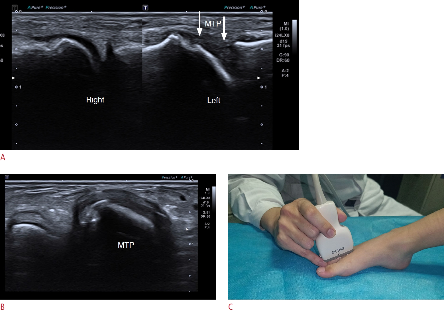
Freiberg’s Disease
Epidemiology
Freiberg’s disease, also referred to as osteochondrosis or avascular necrosis of the second metatarsal head, was first described by Dr Albert H. Freiberg in 1914. It is a rare clinical condition. The disease has an insidious onset and a significant predilection to the adolescent athletic female population [59], and it mostly affects the second metatarsophalangeal (MTP) joint.
Pathophysiology
The etiology of Freiberg’s disease is multifactorial, involving trauma, mechanical arterial compression, genetic susceptibility, and systemic disorders (hypercoagulability, systemic lupus erythematosus, diabetes mellitus, etc.). Nevertheless, the underlying cause of Freiberg’s disease remains obscure.
Clinical Findings
Patients predominantly complain of forefoot chronic pain, swelling, tenderness and restricted motion at the level of the involved MTP joint. The symptoms are exacerbated by walking barefoot or wearing shoes with an elevated heel and alleviated by rest. A sagittal or coronal misaligned pattern of the toes may be evident on a physical examination. The Lachman test should be performed to demonstrate MTP joint instability. Advanced Freiberg’s disease may present with crossover or claw toe deformity [60].
Ultrasound Findings
The US examination to assess the forefoot can be conducted with the patient positioned either supine or sitting. With the patient's knee flexed and feet flat on the bed, the dorsal side of the foot is easily accessible for examination. Alternatively, with the patient's knee extended and leg resting on the bed, both the plantar and dorsal surfaces of the foot can be evaluated without repositioning. This position enables dynamic assessments and facilitates easy comparison with the contralateral foot [61].
On US, the normal cortex of the second metatarsal head presents as a smooth echogenic line accompanied by a reverberation artifact and posterior acoustic shadowing [62].
The US presentation of Freiberg’s disease involves a flattened and fragmented metatarsal head with a rough and irregular bony surface, as well as a swollen adjacent MTP joint [61]. Synovial hyperplasia and anechoic effusions can be observed in the involved joint. Color and power Doppler US may reveal angiogenesis in synovial tissues (Fig. 7) [63].
Hill et al. [64] observed that the earliest radiographic signs of Freiberg’s disease typically appear around 3 to 6 weeks after the initial onset of symptoms. These signs are characterized by subtle widening of the joint space. Although MRI provides superior visualization of stress-related bone marrow edema and parosteal soft tissue changes, high-resolution US has emerged as a primary diagnostic method due to its widespread availability in sports clinic offices and its convenience for on-site physical examinations [62].
Treatment
The initial management is conservative—namely rest, wear modifications, and oral NSAIDs. If these interventions are ineffective, surgical management may be recommended, including debridement, cartilage replacement, osteotomy, implant arthroplasty [65], interpositional arthroplasty, metatarsal head restoration [66], and resection [67,68].
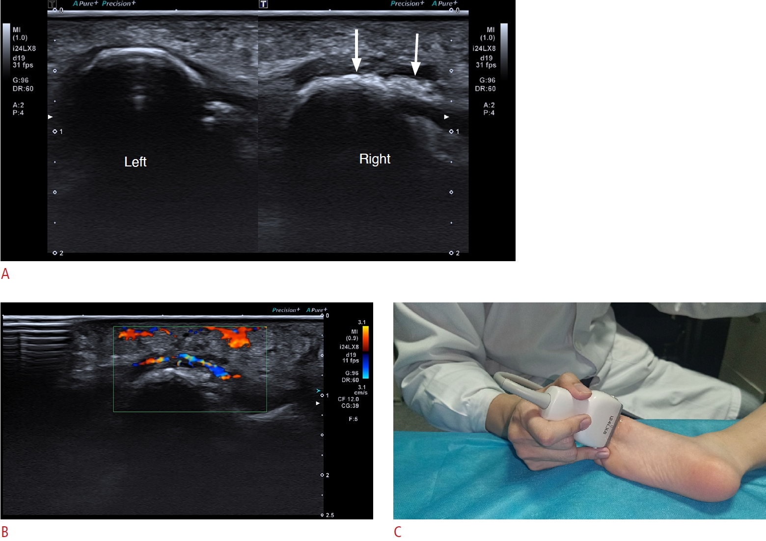
Hallux Sesamoiditis
Epidemiology
There are often two sesamoids lying underneath the first metatarsal head and embedded in the tendon of the flexor hallucis brevis [69]. The tibial sesamoid is more susceptible to injury [70]. Hallux sesamoiditis is a motor impairment caused by repetitive injury to the plantar aspect of the forefoot; it is most often seen in athletic adolescents, such as joggers and dancers [71,72]. During sudden acceleration and deceleration during jumps, the sesamoids undergo excessive impact and load.
Pathophysiology
The hallux sesamoids can absorb weight-bearing pressure, reduce friction, and augment the efficacy of the flexor hallucis brevis tendon. Rapid acceleration and deceleration during jumps can lead to greater shear and axial forces on the sesamoids. Repetitive and excessive stresses eventually cause inflammation and fracture, or even necrosis of the sesamoids [73,74].
Clinical Findings
Hallux sesamoiditis is characterized by pain and tenderness under the first metatarsal head, aggravated by dorsiflexion of the first MTP joint. Patients typically present with an antalgic gait.
Ultrasound Findings
The patient position in a US examination for the presence of hallux sesamoid is the same as that for talocalcaneal coalition.
It is important to correlate the US imaging appearance with physical examination findings. The most common manifestation of hallux sesamoiditis on US is a blurred, rough, and uneven bone cortex, with or without disruption of the sesamoid [75]. The volume of an injured sesamoid may become larger due to compression and inflammation. US may also show thickening of the hallux short flexor tendon with increased vascularity in it and edema of the surrounding soft tissues (Fig. 8). It is challenging to differentiate an acute sesamoid fracture from bipartite sesamoid [76]; therefore, a comparison with the contralateral site is crucial.
Radiography is optimal for the initial evaluation of accessory bones. CT is highly effective in identifying fracture lines, while MRI demonstrates sensitivity in detecting abnormalities in marrow signals and surrounding soft tissues [77]. US is well-suited for a dynamic assessment and can also serve as a guide for percutaneous interventions.
Treatment
Hallux sesamoiditis is typically treated conservatively, including rest, ice, weight-bearing in a stiff-soled shoe, anti-inflammatory drugs, activity modification, carbon-fiber footplate, and cast immobilization [78]. If conservative management fails, surgical management involving excision of the affected sesamoid is warranted to repair the remaining tendon and prevent an ensuing hallux valgus deformity [79,80]. Competitive athletes have demonstrated decreased pain, early return to sport, and few postsurgical complications [79].
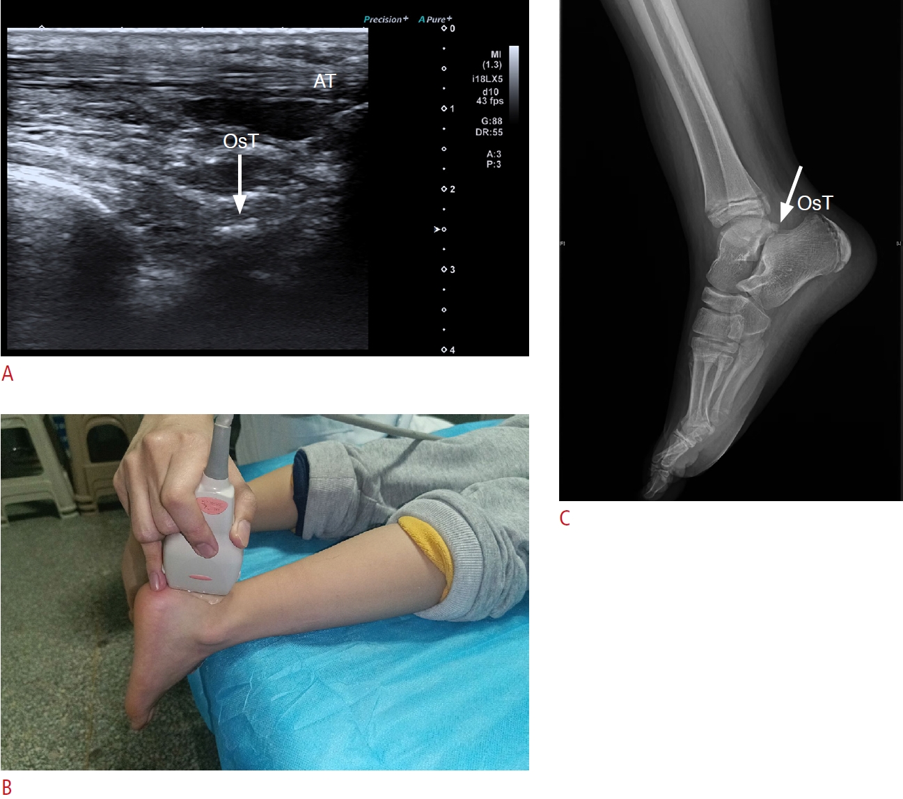
Os Trigonum Syndrome
Epidemiology
Os trigonum is the most frequent accessory ossicle at the ankle. It originates from a failed fusion of the secondary ossification to the posterolateral talar tubercle, replaced with a synchondrosis. Os trigonum may exist in pairs and has triangular, round, or oval morphology. It is easily misdiagnosed as a fracture clinically. Os trigonum syndrome can also be referred to as posterior ankle syndrome and defined as the symptomatic os trigonum from repetitive use or an injury. The prevalence of os trigonum syndrome ranges from 1.7% to 7%. Os trigonum syndrome is predominantly seen in ballet dancers [81], downhill runners, soccer players, and javelin athletes [82] resulting from repeated plantarflexion injuries [83–85].
Pathophysiology
Acute plantar hyperflexion injury and chronic recurrent microtrauma can lead to a fracture of the os trigonum, cartilaginous synchondrosis disruption, or even an avulsion of the posterior talofibular ligament [84,86]. The surrounding posterior soft tissues are compressed between the calcaneus and the posterior talus and secondarily hypertrophied.
Clinical Findings
The symptoms of os trigonum syndrome include chronic pain, stiffness, and swelling of the posterior ankle [84]. Activity limitation is common, particularly while in plantar flexion [86]. A physical examination reveals posterolateral tenderness on palpation. Passive plantar flexion may trigger the pain.
Ultrasound Findings
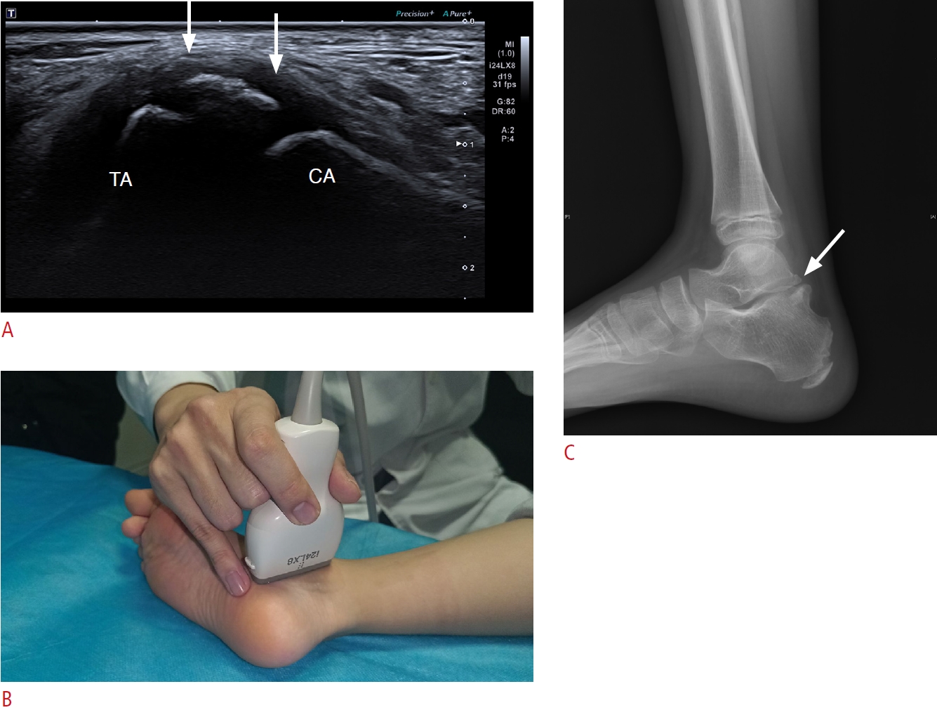
US is typically conducted with the patient lying prone on the examination table, with their feet hanging off the edge.
Os trigonum is located at a distance from the Achilles tendon; therefore, a 9-12 MHz linear array transducer is recommended (Fig. 9) [87].
US of symptomatic os trigonum may show irregular or hypertrophic osseous hyperechogenicity, with a posterior acoustic shadow situated posterior to the talus [76,88,89]. A rough bone cortex and osteophytes of the talus or calcaneus may also be seen. Synovitis in the posterior tibiotalar joint or flexor hallucis longus tenosynovitis may be revealed on a US examination of symptomatic os trigonum [76,89]. MRI offers significant advantages as it can reveal talar edema, providing greater diagnostic clarity [90].
Treatment
Conservative treatments include avoidance of activity, icing, anti-inflammatory medications, physical therapy, and corticosteroid injections. Surgery can be indicated after failure of a 3- to 6-month course of nonsurgical treatment. Os trigonum resection can be performed via an open procedure, posterior endoscopy, or arthroscopy [84,86,91,92].
Conclusion
To utilize US effectively in evaluating foot pain in children and adolescents, radiologists must communicate skillfully with patients, gaining a comprehensive understanding of the pain's location and correlating this information with US findings. Moreover, a thorough comprehension of the abnormal US findings in patients exhibiting typical symptoms will enable radiologists to perform precise and efficient US assessments.


 Download Citation
 PDF Links
 PubReader
 ePub Link
 Full text via DOI


